Alternate header for print version


Licensing
Attribution Non-Commercial; No Derivatives:This image is licensed under a Creative Commons Attribution, Non-Commercial, No Derivatives License. View License Deed | View Legal Code
tweet  
*CIL – Cell Image Library accession number. Please use this to reference an image.

CIL:36007*  Cite 
Description

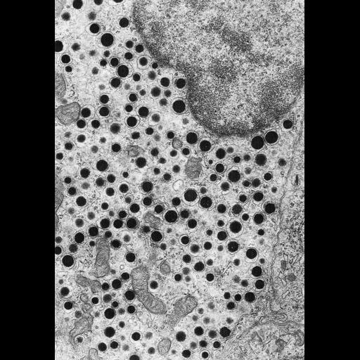
Figure 389 from Chapter 15 (Cytoplasmic Inclusions) of 'The Cell, 2nd Ed.' by Don W. Fawcett M.D. Secretory granules in an alpha cell of an islet of Langerhands in human pancreas. In this EM preparation, the dense core of the secretory vesicle is separated from the membrane, potentially an artifact of specimen preparation. Image by Arthur Like. A PDF copy of the accompanying chapter is available on the ASCB’s BioEDUCATE website.

Biological Sources
NCBI Organism Classification
Homo sapiens
Cell Type
epithelial cell of pancreas
Cellular Component
secretory granule
Biological Context
Biological Process
endocrine process
secretory granule organization
Attribution
Names
Don W. Fawcett
Arthur Like
Published
ISBN 0721635849
Link
http://www.ascb.org/bioeducate/FawcettTheCell...
Citation
Digital Object Identifier (DOI)
doi:10.7295/W9CIL36007
Archival Resource Key (ARK)
ark:/b7295/w9cil36007
Grouping This image is part of a group.
Imaging
Image Type
transmission electron microscopy (TEM)
Image Mode
illumination by electrons
detection of electrons
Parameters Imaged
electron density
Source of Contrast
stain with broad specificity
Visualization Methods
osmium tetroxide
Data Qualifiers
processed data
Dimensions
Spatial Axis Image Size Pixel Size
X 870px ——
Y 1254px ——