Alternate header for print version


Licensing
This image is copyright protected. Any public or private use of this image is subject to prevailing copyright laws. Please contact the content provider of this image for permission requests.
tweet  
*CIL – Cell Image Library accession number. Please use this to reference an image.

CIL:36030*  Cite 
Description

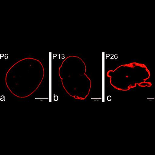
Shown are fibroblasts from a Hutchinson-Gilford Progeria syndrome patient which carries a mutation in the gene for lamin A (red), a major component of the nuclear lamina. The morphology of the envelope shows progressive abnormalities that increase with the number of passages. The passage 6 cell (left) appears much like the wildtype while passage 13 (center) and 26 (right) show nuclear envelope abnormalities. See Fig 1 in R.G. Goldman et al. 2004 Proc Natl Acad Sci 101:8963-8968.

Technical Details

Cells were fixed in formaldehyde or cold methanol, processed for indirect immunofluorescence and images recorded using a Zeiss LSM510 confocal microscope.

Biological Sources
NCBI Organism Classification
Homo sapiens
Cell Type
fibroblast
Cell Line
AG11513
HGADFN003
AG09602B
Cellular Component
nuclear envelope
Attribution
Names
RD Goldman
DK Shumaker
MR Erdos
M Eriksson
AE Goldman
LB Gordon
Y Gruenbaum
S Khuon
M Mendez
R Varga
FS Collins
Published
R.D. Goldman et al. 2004. Proc Natl Acad Sci 101:8963-8968
Pubmed
15184648
Link
nuclear envelope
Citation
Digital Object Identifier (DOI)
doi:10.7295/W9CIL36030
Archival Resource Key (ARK)
ark:/b7295/w9cil36030
Grouping This image is part of a group.
Imaging
Image Type
recorded image
Image Mode
fluorescence microscopy
confocal microscopy
Parameters Imaged
fluorescence emission
Source of Contrast
distribution of epitope
Visualization Methods
Rhodamine
Alexa Fluor 568
Processing History
montage
Data Qualifiers
processed data
Sample Preparation
Methods
formaldehyde fixed tissue
coagulating-fixative fixed tissue
Relation To Intact Cell
whole mounted tissue
Dimensions
Spatial Axis Image Size Pixel Size
X 773px ——
Y 250px ——