Alternate header for print version


Licensing
This image is copyright protected. Any public or private use of this image is subject to prevailing copyright laws. Please contact the content provider of this image for permission requests.
tweet  
*CIL – Cell Image Library accession number. Please use this to reference an image.

CIL:36032*  Cite 
Description

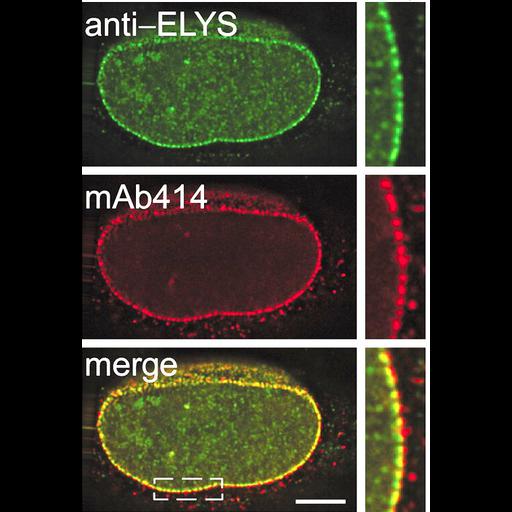
HeLa cells were processed for indirect immunofluorescence with antibodies to two nuclear pore components, ELYS (embryonic large protein derived from yolk sac) and mAb414 against the FG nucleoporins. Both co-localize in a punctate manner to the nuclear envelope as expected for nuclear pore components. See Fig 2A in Rasala et al. Proc Natl Acad Sci 103:17801-17806.

Biological Sources
NCBI Organism Classification
Homo sapiens
Cell Type
epithelial cell
Cell Line
HeLa
Cellular Component
nuclear pore
Biological Context
Biological Process
nuclear pore complex assembly
nucleus organization
Attribution
Names
Beth Rasala
Arturo Orjalo
Zhouxin Shen
Steven Briggs
Douglass Forbes
Published
B Rasala et al. 2006 Proc Natl Acad Sci USA 103:17801-17806
Pubmed
17098863
Link
nuclear envelope
Citation
Digital Object Identifier (DOI)
doi:10.7295/W9CIL36032
Archival Resource Key (ARK)
ark:/b7295/w9cil36032
Imaging
Image Type
recorded image
Image Mode
fluorescence microscopy
3D-deconvolution
Parameters Imaged
fluorescence emission
Source of Contrast
distribution of a specific protein
Processing History
montage
Data Qualifiers
processed data
Sample Preparation
Methods
formaldehyde fixed tissue
Relation To Intact Cell
whole mounted tissue
Dimensions
Spatial Axis Image Size Pixel Size
X 516px ——
Y 759px ——