Alternate header for print version


Licensing
This image is copyright protected. Any public or private use of this image is subject to prevailing copyright laws. Please contact the content provider of this image for permission requests.
tweet  
*CIL – Cell Image Library accession number. Please use this to reference an image.

CIL:36036*  Cite 
Description

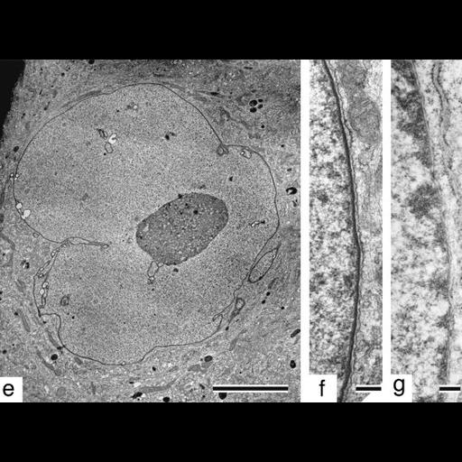
Shown is an image of a thin section showing the ultrastructure of the nucleus and nuclaer envelope in fibroblast from a Hutchinson-Gilford Progeria syndrome patient which carries a mutation in the gene for lamin A, a major component of the nuclear lamina (left and center). The passage 26 cell shows marked abnormalities in the nuclear envelope. Also shown is envelope from a normal fibreblast. Other images in this group show the distribution of other nuclear envelope components in HGPS fibroblasts. See Fig 3 in R.G. Goldman et al. 2004 Proc Natl Acad Sci 101:8963-8968.

Biological Sources
NCBI Organism Classification
Homo sapiens
Cell Type
fibroblast
Cell Line
AG11513
HGADFN003
AG09602B
Cellular Component
nuclear envelope
Attribution
Names
RD Goldman
DK Shumaker
MR Erdos
M Eriksson
AE Goldman
LB Gordon
Y Gruenbaum
S Khuon
M Mendez
R Varga
FS Collins
Published
R.D. Goldman et al. 2004. Proc Natl Acad Sci 101:8963-8968
Pubmed
15184648
Link
nuclear envelope
Citation
Digital Object Identifier (DOI)
doi:10.7295/W9CIL36036
Archival Resource Key (ARK)
ark:/b7295/w9cil36036
Grouping This image is part of a group.
Imaging
Image Type
recorded image
Image Mode
transmission electron microscopy (TEM)
Parameters Imaged
electron density
Source of Contrast
differences in adsorption or binding of stain
Visualization Methods
osmium tetroxide
uranyl salt
Processing History
montage
Data Qualifiers
processed data
Sample Preparation
Methods
glutaraldehyde fixed tissue
tissue in plastic embedment
Relation To Intact Cell
microtome-sectioned tissue
Dimensions
Spatial Axis Image Size Pixel Size
X 547px ——
Y 406px ——