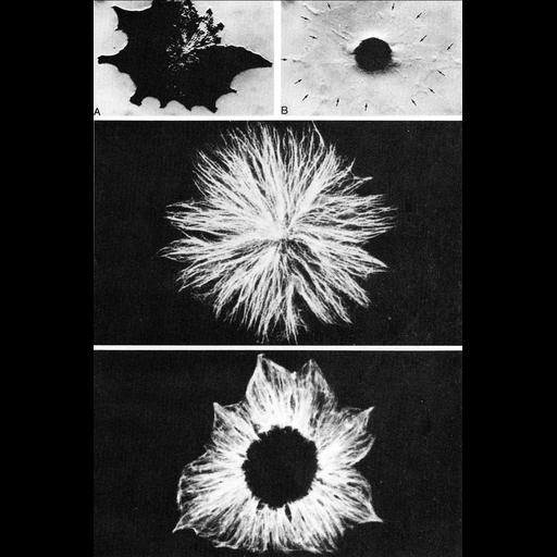
Figures 404 (upper panels A, B), 405 (middle) and 406 (lower) from Chapter 16 (Cytoplasmic matrix and cytoskeleton) of 'The Cell, 2nd Ed.' by Don W. Fawcett M.D. Microtubules and pigment granule transport. Upper panels compare dispersed (A) vs. aggregated (B) pigment granule observed using interference microscopy in living fish melanophores. Arrows in B show that the outer boundary of the cell remains unchanged. Middle panel shows radially oriented microtubules in a melanophore with dispersed pigment, revealed by antibody staining of tubulin with FITC-conjugated secondary. In cells with aggregated pigment, the radial disposition remains unchanged, although in the region of concentrated pigments, only a few fibers can be observed (lower panel). Figures from Schliwa, Osborn and Weber, J. Cell Biol. 76:229-236, 1978 (PMID:338618). A PDF copy of the accompanying chapter is available on the ASCB’s BioEDUCATE website.
| Spatial Axis | Image Size | Pixel Size |
|---|---|---|
| X | 861px | —— |
| Y | 1301px | —— |