Alternate header for print version


Licensing
Attribution Non-Commercial; No Derivatives:This image is licensed under a Creative Commons Attribution, Non-Commercial, No Derivatives License. View License Deed | View Legal Code
tweet  
*CIL – Cell Image Library accession number. Please use this to reference an image.

CIL:36059*  Cite 
Description

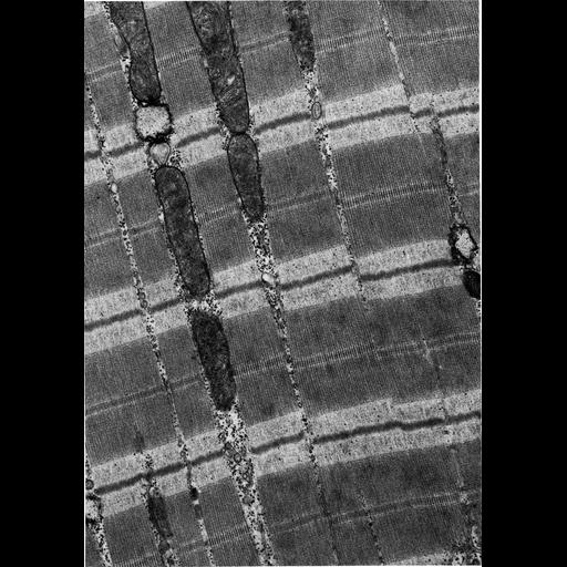
Figure 427 from Chapter 16 (Cytoplasmic matrix and cytoskeleton) of 'The Cell, 2nd Ed.' by Don W. Fawcett M.D. Papillary muscle of cat heart shows the cross-banded organization of striated muscle. Myosin filaments compose the uniform dark A bands. The lighter I bands of actin filaments are bisected by the dense Z line. A PDF copy of the accompanying chapter is available on the ASCB’s BioEDUCATE website.

Biological Sources
NCBI Organism Classification
Felis catus
Cell Type
cardiac muscle cell
Cellular Component
actomyosin, actin part
actomyosin, myosin complex part
A band
I band
Z disc
Biological Context
Biological Process
regulation of cardiac muscle contraction
Attribution
Names
Don W. Fawcett
Published
ISBN 0721635849
Link
http://www.ascb.org/bioeducate/FawcettTheCell...
Citation
Digital Object Identifier (DOI)
doi:10.7295/W9CIL36059
Archival Resource Key (ARK)
ark:/b7295/w9cil36059
Grouping This image is part of a group.
Imaging
Image Type
transmission electron microscopy (TEM)
Image Mode
illumination by electrons
detection of electrons
Parameters Imaged
electron density
Source of Contrast
stain with broad specificity
Visualization Methods
osmium tetroxide
Data Qualifiers
processed data
Dimensions
Spatial Axis Image Size Pixel Size
X 892px ——
Y 1273px ——