Alternate header for print version


Licensing
Attribution Non-Commercial; No Derivatives:This image is licensed under a Creative Commons Attribution, Non-Commercial, No Derivatives License. View License Deed | View Legal Code
tweet  
*CIL – Cell Image Library accession number. Please use this to reference an image.

CIL:36060*  Cite 
Description

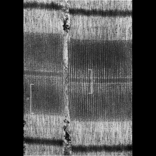
Figure 428 from Chapter 16 (Cytoplasmic matrix and cytoskeleton) of 'The Cell, 2nd Ed.' by Don W. Fawcett M.D. Papillary muscle of cat heart shows one sarcomere length of two adjacent myofibrils at high magnification. The darker A bands are composed of a parallel array of myosin filaments, about 10-11 nm thick and about 1.5 µm long. The lighter I bands of actin filaments are about 6 nm thick extending from the dense central Z line. The bracket labeled X indicates the region of overlap of the two sets of interdigitating filaments. The bracket labeled Y indicates the central region of the A band, composed of only myosin filaments. A PDF copy of the accompanying chapter is available on the ASCB’s BioEDUCATE website.

Biological Sources
NCBI Organism Classification
Felis catus
Cell Type
cardiac muscle cell
Cellular Component
actomyosin, actin part
actomyosin, myosin complex part
A band
I band
Z disc
Biological Context
Biological Process
regulation of cardiac muscle contraction
Attribution
Names
Don W. Fawcett
Published
ISBN 0721635849
Link
http://www.ascb.org/bioeducate/FawcettTheCell...
Citation
Digital Object Identifier (DOI)
doi:10.7295/W9CIL36060
Archival Resource Key (ARK)
ark:/b7295/w9cil36060
Grouping This image is part of a group.
Imaging
Image Type
transmission electron microscopy (TEM)
Image Mode
illumination by electrons
detection of electrons
Parameters Imaged
electron density
Source of Contrast
stain with broad specificity
Visualization Methods
osmium tetroxide
Data Qualifiers
processed data
Dimensions
Spatial Axis Image Size Pixel Size
X 891px ——
Y 1276px ——