Alternate header for print version


Licensing
Public Domain: This image is in the public domain and thus free of any copyright restrictions. However, as is the norm in scientific publishing and as a matter of courtesy, any user should credit the content provider for any public or private use of this image whenever possible. Learn more
tweet  
*CIL – Cell Image Library accession number. Please use this to reference an image.

CIL:36594*  Cite 
Description

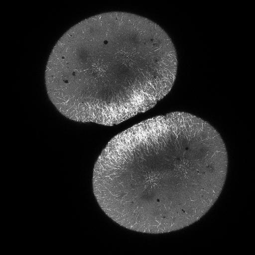
4D video of zebrafish embryo mitosis and cytokinesis. This time lapse video of z-series images was taken of a zebrafish embryo expressing EMTB-3XGFP (a microtubule marker) taken shortly after fertilization.

Technical Details

Images were collected on a Zeiss LSM 710 with a 20X 1.0 NA objective.

Biological Sources
NCBI Organism Classification
Danio rerio
Cell Type
embryo
Cellular Component
microtubule
spindle
aster
Biological Context
Biological Process
mitosis
cytokinesis after mitosis
Attribution
Names
Martin Wuehr
Timothy Mitchison
Citation
Digital Object Identifier (DOI)
doi:10.7295/W9CIL36594
Archival Resource Key (ARK)
ark:/b7295/w9cil36594
Imaging
Image Type
recorded image
Image Mode
single-spot confocal microscopy
Parameters Imaged
fluorescence emission
Source of Contrast
distribution of a specific protein
Visualization Methods
EGFP
Processing History
unprocessed raw data
Data Qualifiers
raw, unprocessed data
suitable for spatial measurements
Sample Preparation
Methods
living tissue
Relation To Intact Cell
whole mounted tissue
Dimensions
Spatial Axis Image Size Pixel Size
X 2048px 0.2595µm
Y 2048px 0.2595µm
Z 10px 4.915µm