Alternate header for print version


Licensing
Public Domain: This image is in the public domain and thus free of any copyright restrictions. However, as is the norm in scientific publishing and as a matter of courtesy, any user should credit the content provider for any public or private use of this image whenever possible. Learn more
tweet  
*CIL – Cell Image Library accession number. Please use this to reference an image.

CIL:36665*  Cite 
Description

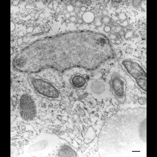
The first culture of P. multimicronucleatum which I had obtained from Carolina Biological Supply House contained a bacterial endosymbiont that resided specifically in the space between the two membranes of the nuclear envelope or in outpocketings of this space so that the bacteria were enclosed by the outer membrane of the nuclear envelope. Such endosymbionts have been observed before (Jenkins, J. Gen. Microbiol. 61:355-359, 1970). In this micrograph the bacteria are associated with the micronucleus, which has a remnant bundle of fibers and microtubules, but bacteria are also associated with the macronucleus (not shown). TEM taken on 12/9/70 by R. Allen with Hitachi HU11A operating at 75kV. Neg. 19,500X. Bar = 0.2µm.

Technical Details

Standard glutaraldehyde fixation followed by osmium tetroxide, dehydrated in alcohol and embedded in an epoxy resin. Microtome sections prepared at approximately 75nm thickness. The negative was printed to paper and the image was scanned to Photoshop. This digitized image is available for qualitative analysis. Additional information available at (http://www5.pbrc.hawaii.edu/allen/).

Biological Sources
NCBI Organism Classification
Paramecium multimicronucleatum
Cell Type
cell by organism
eukaryotic cell
Eukaryotic Protist
Ciliated Protist
Cellular Component
symbiont-containing vacuole membrane
micronucleus
Biological Context
Biological Process
detection of symbiont
micronucleus organization
Attribution
Names
Richard Allen (University of Hawaii)
Citation
Digital Object Identifier (DOI)
doi:10.7295/W9CIL36665
Archival Resource Key (ARK)
ark:/b7295/w9cil36665
Imaging
Image Type
transmission electron microscopy (TEM)
illumination by electrons
Image Mode
detection of electrons
film
Parameters Imaged
electron density
Source of Contrast
stain with broad specificity
Visualization Methods
stain with broad specificity
uranyl salt
lead salt
osmium tetroxide
Processing History
recorded image
film
Print from negative scanned for Photoshop.
Data Qualifiers
processed data
Dimensions
Spatial Axis Image Size Pixel Size
X 2600px ——
Y 3000px ——