Alternate header for print version


Licensing
Attribution Non-Commercial Share Alike:This image is licensed under a Creative Commons Attribution, Non-Commercial Share Alike License. View License Deed | View Legal Code
tweet  
*CIL – Cell Image Library accession number. Please use this to reference an image.

CIL:37149*  Cite 
Description

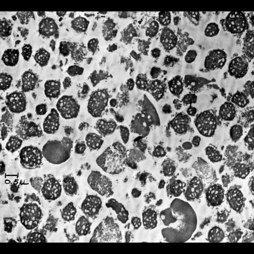
Transmission electron micrograph of the nucleolar fraction from a cell fractionation of guinea pig liver. Image made available by James D. Jamieson and the Department of Cell Biology, Yale University School of Medicine.

Technical Details

Original 3.25 in. x 4 in. lantern slides were scanned at 600dpi.

Biological Sources
NCBI Organism Classification
Cavia porcellus
Cell Type
liver cell
Cellular Component
nucleolus
Attribution
Names
George E. Palade
R. Maggio
Link
George E. Palade EM Slide Collection
Citation
Digital Object Identifier (DOI)
doi:10.7295/W9CIL37149
Archival Resource Key (ARK)
ark:/b7295/w9cil37149
Grouping This image is part of a group.
Sample Preparation
Methods
cell fractionation
Relation To Intact Cell
sectioned tissue
Dimensions
Spatial Axis Image Size Pixel Size
X 6000px ——
Y 5461px ——