Alternate header for print version


Licensing
Attribution Non-Commercial Share Alike:This image is licensed under a Creative Commons Attribution, Non-Commercial Share Alike License. View License Deed | View Legal Code
tweet  
*CIL – Cell Image Library accession number. Please use this to reference an image.

CIL:37152*  Cite 
Description

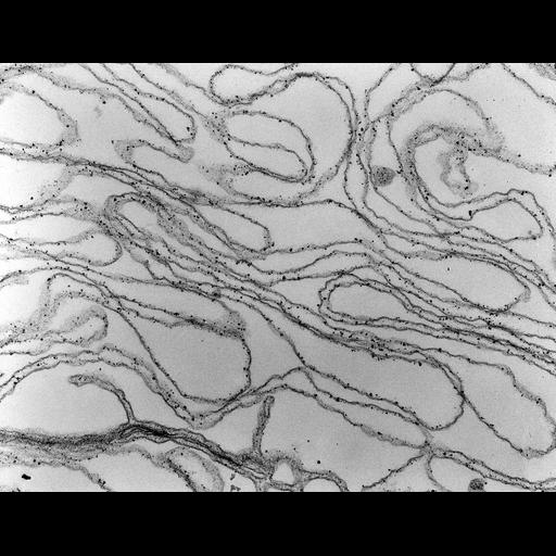
Transmission electron micrograph of spectrin in cell fractionation. Dr. V. T. Marchesi and colleagues took advantage of the high purity of red cell ghosts obtained by hypotonic lysis to analyze the properties of membrane proteins. They identified spectrin as a major red cell membrane cytoskeletal protein responsible for maintaining the biconcave shape of red cells. Image made available by James D. Jamieson and the Department of Cell Biology, Yale University School of Medicine.

Technical Details

Original 3.25 in. x 4 in. lantern slides were scanned at 600dpi. Original Magnification: x25,000. Relevant citations: Marchesi, V.T. and G.E. Palade. 1967. J. Cell Biol. 35:385-404. Nicoloson, G.L., V.T. Marchesi and S.J. Singer. 1971. J. Cell Biol. 51:265-272. Marchesi, V.T. and E. Steers, Jr. 1968. Science. 159:203-204.

Biological Sources
Cell Type
blood cell
red blood cell
Cellular Component
plasma membrane
spectrin
Attribution
Names
George E. Palade
Vincent. T. Marchesi
Link
George E. Paldae EM Slide Collection
Citation
Digital Object Identifier (DOI)
doi:10.7295/W9CIL37152
Archival Resource Key (ARK)
ark:/b7295/w9cil37152
Grouping This image is part of a group.
Imaging
Image Type
recorded image
Image Mode
transmission electron microscopy (TEM)
Parameters Imaged
electron density
Source of Contrast
differences in adsorption or binding of stain
Visualization Methods
ferratin conjugated antibody
Sample Preparation
Methods
hypotonic lysis
Relation To Intact Cell
sectioned tissue
Dimensions
Spatial Axis Image Size Pixel Size
X 6000px ——
Y 4630px ——