Alternate header for print version


Licensing
Attribution Only: This image is licensed under a Creative Commons Attribution License. View License Deed | View Legal Code
tweet  
*CIL – Cell Image Library accession number. Please use this to reference an image.

CIL:37157*  Cite 
Description

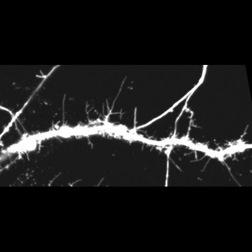
This is Video S2 which corresponds to still images in top row of Figure 2D. It shows the spine dynamics of control neurons. Time-lapse confocal imaging was performed on DIV14 hippocampal neurons co-expressing GFP. Images were acquired every 1-minute using confocal microscopy. 3 frames/sec shown.

Technical Details

Confocal images were collected on an Olympus Fluoview 1000 microscope (IX81 base) equipped with a 60X/1.35 NA (oil)UPLSAPO 60X objective (Olympus). GFP was excited using the 488 nm laser line of a multi Ar laser. Fluorescence emission was collected using the following dichroic mirror/filter: SDM560/BA505–525 (GFP).

Biological Sources
NCBI Organism Classification
Rattus
Cell Type
CNS neuron (sensu Vertebrata)
Cellular Component
dendritic spine
dendrite
dendritic shaft
cytoplasm
Attribution
Names
Jennifer L. Hodges
Karen Newell-Litwa
Hannelore Asmussen
Miguel Vicente-Manzanares
Alan Rick Horwitz
Link
http://www.plosone.org/article/info%3Adoi%2F1...
Citation
Digital Object Identifier (DOI)
doi:10.7295/W9CIL37157
Archival Resource Key (ARK)
ark:/b7295/w9cil37157
Grouping This image is part of a group.
Imaging
Image Type
time lapse microscopy
Image Mode
confocal microscopy
Parameters Imaged
fluorescence emission
Source of Contrast
distribution of a specific protein
Visualization Methods
EGFP
Processing History
unprocessed raw data
Data Qualifiers
raw, unprocessed data
suitable for spatial measurements
Sample Preparation
Methods
living tissue
Relation To Intact Cell
dispersed cells in vitro
Dimensions
Spatial Axis Image Size Pixel Size
X 759px 0.06µm
Y 367px 0.06µm
Time 60 seconds 20