Alternate header for print version


Licensing
Attribution Non-Commercial Share Alike:This image is licensed under a Creative Commons Attribution, Non-Commercial Share Alike License. View License Deed | View Legal Code
tweet  
*CIL – Cell Image Library accession number. Please use this to reference an image.

CIL:37185*  Cite 
Description

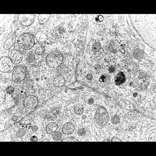
Transmission electron micrograph of the junctional complexes at the intersection of three hepatocytes. This image was taken from liver tissues from an ethanol fed rat and also shows the bile canaliculus. Smooth endoplasmic reticulum and associated vesicles are prominent. Image made available by James D. Jamieson and the Department of Cell Biology, Yale University School of Medicine.

Technical Details

Original 3.25 in. x 4 in. lantern slides were scanned at 600dpi. Original Magnification: x10,050.

Biological Sources
NCBI Organism Classification
Rattus
Cell Type
hepatocyte
Cellular Component
cell-cell junction
mitochondrion
nucleus
Golgi apparatus
smooth endoplasmic reticulum
rough endoplasmic reticulum
Biological Context
Biological Process
cell-cell junction organization
Mouse Gross Anatomy
bile canaliculus
Citation
Digital Object Identifier (DOI)
doi:10.7295/W9CIL37185
Archival Resource Key (ARK)
ark:/b7295/w9cil37185
Grouping This image is part of a group.
Imaging
Image Type
recorded image
Image Mode
transmission electron microscopy (TEM)
Parameters Imaged
electron density
Source of Contrast
differences in adsorption or binding of stain
Visualization Methods
osmium tetroxide
Processing History
unprocessed raw data
Sample Preparation
Methods
chemically fixed tissue
Relation To Intact Cell
microtome-sectioned tissue
Dimensions
Spatial Axis Image Size Pixel Size
X 6000px ——
Y 4913px ——