Alternate header for print version


Licensing
Attribution Non-Commercial Share Alike:This image is licensed under a Creative Commons Attribution, Non-Commercial Share Alike License. View License Deed | View Legal Code
tweet  
*CIL – Cell Image Library accession number. Please use this to reference an image.

CIL:37205*  Cite 
Description

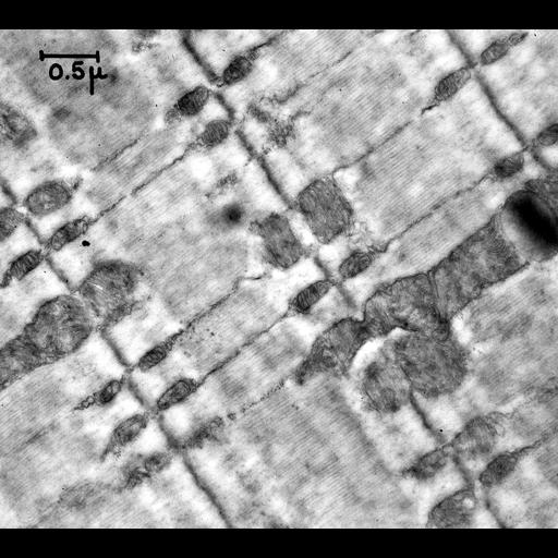
Transmission electron micrograph of rat sartorius striated muscle showing the regular arrangement of the actin-myosin myofilaments and abundant mitochondria. This 1956 TEM image has historical value as one of the earliest views of muscle ultrastructure. Image made available by James D. Jamieson and the Department of Cell Biology, Yale University School of Medicine.

Technical Details

Additional reference: Porter, K.R. and G.E. Palade. 1957. Studies on the endoplasmic reticulum: Its form and distribution in striated muscle. J. Biophys. Biochem. Cytol. 3:269-300. Original 3.25 in. x 4 in. lantern slides were scanned at 600dpi.

Biological Sources
NCBI Organism Classification
Rattus
Cell Type
sartorius muscle
Cellular Component
mitochondrion
sarcoplasmic reticulum
muscle myosin complex
myofibril
Biological Context
Biological Process
voluntary skeletal muscle contraction
Attribution
Names
George E. Palade
Keith Porter
Link
George E. Palade EM Slide Collection
Citation
Digital Object Identifier (DOI)
doi:10.7295/W9CIL37205
Archival Resource Key (ARK)
ark:/b7295/w9cil37205
Grouping This image is part of a group.
Imaging
Image Type
recorded image
Image Mode
transmission electron microscopy (TEM)
Parameters Imaged
electron density
Source of Contrast
differences in adsorption or binding of stain
Visualization Methods
osmium tetroxide
Processing History
unprocessed raw data
Sample Preparation
Methods
chemically fixed tissue
Relation To Intact Cell
sectioned tissue
Dimensions
Spatial Axis Image Size Pixel Size
X 6000px ——
Y 5377px ——