Alternate header for print version


Licensing
Attribution Non-Commercial Share Alike:This image is licensed under a Creative Commons Attribution, Non-Commercial Share Alike License. View License Deed | View Legal Code
tweet  
*CIL – Cell Image Library accession number. Please use this to reference an image.

CIL:37227*  Cite 
Description

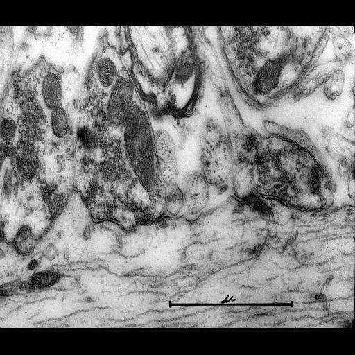
Transmission electron micrograph of nerve cell terminals on medulla dendrites. The body of the dendrite (lower part of image) contains aligned microtubules (neurotubule). Synapses are seem on the dendrite membrane. Note that this very early (1954) micrograph is primarily of historical interest since it pre-dates glutaraldehye fixation which was introduced in the early 60's and is considered essential for adequate nerve tissue preservation. Examples of nerve tissue micrographs prepared in 1965 are CIL:37218, CIL:37219 and CIL:37220. Image made available by James D. Jamieson and the Department of Cell Biology, Yale University School of Medicine.

Technical Details

Original 3.25 in. x 4 in. lantern slides were scanned at 600dpi. Origianl magnification x28,000.

Biological Sources
Cell Type
neuron
Cellular Component
dendrite
synapse
microtubule
Biological Context
Biological Process
transmission of nerve impulse
Human Development Anatomy
medulla
Attribution
Names
George E. Palade
Link
George E. Palade EM Slide Collection
Citation
Digital Object Identifier (DOI)
doi:10.7295/W9CIL37227
Archival Resource Key (ARK)
ark:/b7295/w9cil37227
Grouping This image is part of a group.
Imaging
Image Type
recorded image
Image Mode
transmission electron microscopy (TEM)
Parameters Imaged
electron density
Source of Contrast
differences in adsorption or binding of stain
Visualization Methods
osmium tetroxide
Processing History
unprocessed raw data
Sample Preparation
Methods
chemically fixed tissue
Relation To Intact Cell
sectioned tissue
Dimensions
Spatial Axis Image Size Pixel Size
X 6000px ——
Y 5106px ——