Alternate header for print version


Licensing
Attribution Non-Commercial Share Alike:This image is licensed under a Creative Commons Attribution, Non-Commercial Share Alike License. View License Deed | View Legal Code
tweet  
*CIL – Cell Image Library accession number. Please use this to reference an image.

CIL:37241*  Cite 
Description

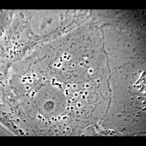
This phase contrast image of a fibroblast was made available by James D. Jamieson and the Department of Cell Biology, Yale University School of Medicine.

Technical Details

Original 3.25 in. x 4 in. lantern slides were scanned at 600dpi. The original magnification was 1,000X.

Biological Sources
Cell Type
fibroblast
Cellular Component
nucleus
plasma membrane
mitochondrion
Attribution
Names
Keith Porter
Link
George E. Palade EM Slide Collection
Citation
Digital Object Identifier (DOI)
doi:10.7295/W9CIL37241
Archival Resource Key (ARK)
ark:/b7295/w9cil37241
Grouping This image is part of a group.
Dimensions
Spatial Axis Image Size Pixel Size
X 1849px ——
Y 1610px ——