Alternate header for print version


Licensing
Attribution Non-Commercial Share Alike:This image is licensed under a Creative Commons Attribution, Non-Commercial Share Alike License. View License Deed | View Legal Code
tweet  
*CIL – Cell Image Library accession number. Please use this to reference an image.

CIL:37257*  Cite 
Description

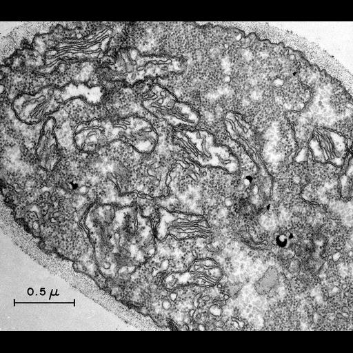
An electron micrograph of a thin section of a hypha of the fungal ascomycete Neurospora crassa. The cell wall, mitochondria and cytoplasmic ribosomes are prominent. Image made available by James D. Jamieson and the Department of Cell Biology, Yale University School of Medicine.

Technical Details

Reference Luck, D.J.L. 1963. Formation of mitochondria in Neurospora crassa: A quantitative radioautographic study. J. Cell Biol. 16:483-499. Original 3.25 in. x 4 in. lantern slides were scanned at 600dpi. Original magnification 20,000X.

Biological Sources
NCBI Organism Classification
Neurospora crassa
Cellular Component
mitochondrion
cell wall
ribosome
Biological Context
Biological Process
organelle organization
Attribution
Names
D. Luck
Link
George E. Palade EM Slide Collection
Citation
Digital Object Identifier (DOI)
doi:10.7295/W9CIL37257
Archival Resource Key (ARK)
ark:/b7295/w9cil37257
Grouping This image is part of a group.
Imaging
Image Type
recorded image
Image Mode
transmission electron microscopy (TEM)
Parameters Imaged
electron density
Source of Contrast
differences in adsorption or binding of stain
Processing History
unprocessed raw data
Sample Preparation
Methods
chemically fixed tissue
tissue in plastic embedment
Relation To Intact Cell
sectioned tissue
Dimensions
Spatial Axis Image Size Pixel Size
X 6000px ——
Y 5262px ——