Alternate header for print version


Licensing
Attribution Non-Commercial Share Alike:This image is licensed under a Creative Commons Attribution, Non-Commercial Share Alike License. View License Deed | View Legal Code
tweet  
*CIL – Cell Image Library accession number. Please use this to reference an image.

CIL:38049*  Cite 
Description

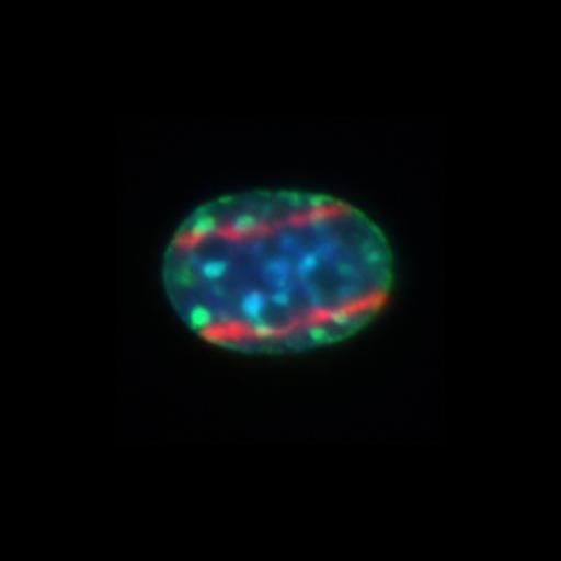
3T3 cells, 5min after the induction of DNA damage by microirradiation with a 405 laser. Cells were immunostained with antibody against H3K9me3 (green), a post-translational histone modification enriched at the pericentric heterochromatin domains. DNA damage sites (sites of irradiation) were visualized indirectly by H2AX (red) staining, DNA is stained with Hoescht 33258 (blue). This image is part of the original data set contributing to Baldeyron et al. (2011) HP1α recruitment to DNA damage by p150CAF-1 promotes homologous recombination repair J. Cell Biol. 193: 81-95.

Biological Sources
NCBI Organism Classification
Mus musculus
Cell Line
3T3-Swiss albino
Cellular Component
nucleus
heterochromatin
Biological Context
Biological Process
regulation of DNA repair
Attribution
Names
Celine Baldeyron
Gaston Soria
Daniele Roche
Adam J. L. Cook
Genevieve Almouzni
Link
http://jcb-dataviewer.rupress.org/jcb/browse/...
Citation
Digital Object Identifier (DOI)
doi:10.7295/W9CIL38049
Archival Resource Key (ARK)
ark:/b7295/w9cil38049
Imaging
Image Type
recorded image
Image Mode
fluorescence microscopy
Parameters Imaged
fluorescence emission
Source of Contrast
distribution of epitope
differences in adsorption or binding of stain
Visualization Methods
Hoechst 33258
Alexa Fluor 488
Alexa Fluor 594
Data Qualifiers
processed data
Sample Preparation
Methods
formaldehyde fixed tissue
detergent permeabilized
Relation To Intact Cell
dispersed cells in vitro
Dimensions
Spatial Axis Image Size Pixel Size
X 300px ——
X 300px ——