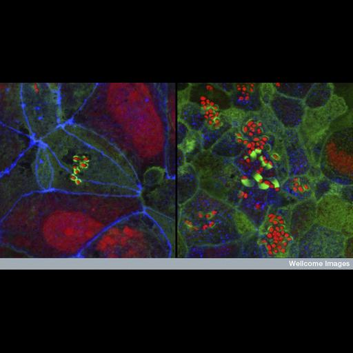
This pair of confocal micrographs demonstrates how a disease-causing strain of E. coli bacteria brings about diarrhoea by breaking down the waterproof barriers between the cells. The bacteria are seen as small red dots attached to the surface of intestinal cells making tiny pedestals out of one of the cell's own proteins (bright green). Once attached, the bacteria send signals into the cells, causing the tight junctions (blue) between the cells to break down. Water is then able to seep out between the cells into the intestine, leading to diarrhoea. The image on the left shows an early stage in the process where the tight junctions are still intact and show as continuous blue lines between the cells. The image on the right is taken later in the process, once the tight junctions have broken down. Their remnants appear as blue dots.
B0006873 Confocal micrograph 2007 Collection: Medical Photographic Library. Copyrighted work available under Creative Commons by-nc-nd 2.0 UK: England & Wales, see http://creativecommons.org/licenses/by-nc-nd/2.0/uk/
| Spatial Axis | Image Size | Pixel Size |
|---|---|---|
| X | 800px | —— |
| Y | 424px | —— |