Alternate header for print version


Licensing
Attribution Non-Commercial; No Derivatives:This image is licensed under a Creative Commons Attribution, Non-Commercial, No Derivatives License. View License Deed | View Legal Code
tweet  
*CIL – Cell Image Library accession number. Please use this to reference an image.

CIL:38918*  Cite 
Description

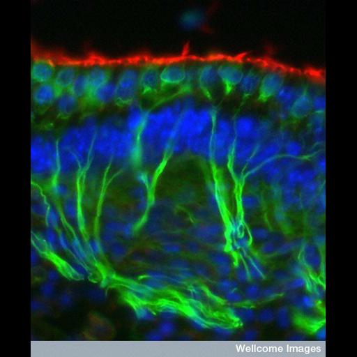
Confocal image of a section through a region of the inner ear called the vestibular organ, which is part of a complex arrangement of tubes and chambers that work together to enable us to keep our balance. As our heads move, fluid flows over the hair bundles (red) pushing them backwards and forwards. The nerves (green) connected to the base of the hair cells then send messages to the brain. By interpreting these movements the brain enables us to make any compensating movements and so keep our balance. The cell nuclei are stained blue.

Technical Details

B0005954 Nerves and hair cells in the organ of balance. Wellcome Images available under the following creative commons usage http://creativecommons.org/licenses/by-nc-nd/2.0/uk/

Biological Sources
Cell Type
vestibular hair cell
neuron
Cellular Component
nucleus
Attribution
Names
Dawn Davies
Link
Wellcome Images
Citation
Digital Object Identifier (DOI)
doi:10.7295/W9CIL38918
Archival Resource Key (ARK)
ark:/b7295/w9cil38918
Imaging
Image Type
recorded image
Image Mode
confocal microscopy
Parameters Imaged
fluorescence emission
Source of Contrast
distribution of epitope
Visualization Methods
primary antibody plus labeled secondary antibody
Processing History
unprocessed raw data
Sample Preparation
Methods
chemically fixed tissue
Relation To Intact Cell
sectioned tissue
Dimensions
Spatial Axis Image Size Pixel Size
X 476px ——
Y 576px ——