Alternate header for print version


Licensing
Attribution Non-Commercial; No Derivatives:This image is licensed under a Creative Commons Attribution, Non-Commercial, No Derivatives License. View License Deed | View Legal Code
tweet  
*CIL – Cell Image Library accession number. Please use this to reference an image.

CIL:38950*  Cite 
Description

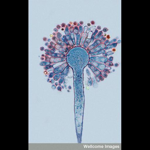
Phase contrast micrograph of spore formation and release in Aspergillus, an ascomycete, using a semi-thin stained section stained blue. Chains of asexual spores (conidia) bud off from the projections (phialides) on special hyphae called conidiophores, one of which is shown here, which in Aspergillus terminate in a characteristic 'mop head' . Most Aspergillus species are saprophytes, feeding on on decaying organic matter. Some species produce mycotoxins that can bulid up in livestock or humans that eat infected foodstuffs. Some are respiratory tract pathogens, causing lung diseases in humans, poultry and other animals when the spores are inhaled, for example, farmer's lung or aspergillosus. Fermentation by certain Aspergillus species is involved in the production of soy sauce.

Technical Details

B0004539 Aspergillus spore formation (conidia), phase contrast. Wellcome Images available under the following creative commons usage http://creativecommons.org/licenses/by-nc-nd/2.0/uk/

Biological Sources
NCBI Organism Classification
Agromyces sp. R
Cell Type
fungal asexual spore
hyphal cell
conidiophore
Cellular Component
hyphal tip
phialides
Biological Context
Biological Process
asexual sporulation
Attribution
Names
Spike Walker
Link
Wellcome Images
Citation
Digital Object Identifier (DOI)
doi:10.7295/W9CIL38950
Archival Resource Key (ARK)
ark:/b7295/w9cil38950
Imaging
Image Type
recorded image
Image Mode
phase contrast microscopy
Parameters Imaged
elastic scattering of photons
Source of Contrast
differences in amount of elastic light scattering
differences in adsorption or binding of stain
Visualization Methods
blue stain
Processing History
unprocessed raw data
Sample Preparation
Relation To Intact Cell
sectioned tissue
Dimensions
Spatial Axis Image Size Pixel Size
X 357px ——
Y 576px ——