Alternate header for print version


Licensing
Attribution Non-Commercial; No Derivatives:This image is licensed under a Creative Commons Attribution, Non-Commercial, No Derivatives License. View License Deed | View Legal Code
tweet  
*CIL – Cell Image Library accession number. Please use this to reference an image.

CIL:38960*  Cite 
Description

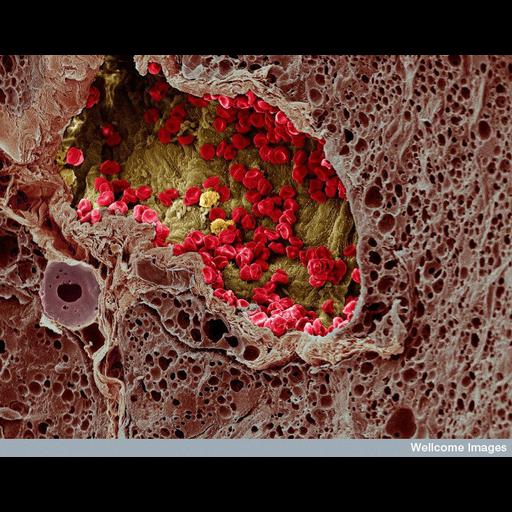
A color-enhanced, freeze-fracture scanning electron micrograph of a blood vessel that has grown into a melanoma and is providing nourishment to it. Numerous red blood cells and three white blood cells can be seen within the blood vessel.

Technical Details

B0003655 SEM of blood vessel in a melanoma - colored. Wellcome Images available under the following creative commons usage http://creativecommons.org/licenses/by-nc-nd/2.0/uk/

Biological Sources
Cell Type
blood cell
Cellular Component
cell surface
Biological Context
Biological Process
angiogenesis
Human Disease
melanoma
Attribution
Names
K. Hodivala-Dilke
M. Stone
Link
Wellcome Images
Citation
Digital Object Identifier (DOI)
doi:10.7295/W9CIL38960
Archival Resource Key (ARK)
ark:/b7295/w9cil38960
Imaging
Image Type
recorded image
Image Mode
scanning electron microscopy (SEM)
Parameters Imaged
elastic scattering of electrons
Source of Contrast
differences in deposition of metal shadow
Visualization Methods
visualization of contiguous regions
Processing History
colorized
Sample Preparation
Methods
freeze_fracture/freeze_etch
Relation To Intact Cell
sec
Dimensions
Spatial Axis Image Size Pixel Size
X 733px ——
Y 576px ——