Alternate header for print version


Licensing
Attribution Non-Commercial; No Derivatives:This image is licensed under a Creative Commons Attribution, Non-Commercial, No Derivatives License. View License Deed | View Legal Code
tweet  
*CIL – Cell Image Library accession number. Please use this to reference an image.

CIL:38978*  Cite 
Description

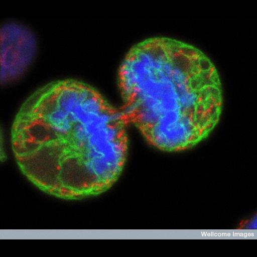
Human melanoma cell undergoing cell division. The chromosomes (blue) have separated and the two daughter cells have almost split apart - only a small bridge of cytoplasm remains. The green staining labels the endoplasmic reticulum and the red labels the mitochondria. The image was produced on a confocal microscope; the ER and mitochondria are from a single optical section but the chromosomes are a 3D reconstruction from a series of sections.

Technical Details

B0003294 Human melanoma cell dividing. Wellcome Images available under the following creative commons usage http://creativecommons.org/licenses/by-nc-nd/2.0/uk/

Biological Sources
NCBI Organism Classification
Homo sapiens
Cell Type
melanocyte
Cellular Component
endoplasmic reticulum
condensed chromosome
mitochondrion
Biological Context
Biological Process
mitosis
Human Disease
melanoma
Attribution
Names
Paul J. Smith
Rachel Errington
Link
Wellcome Images
Citation
Digital Object Identifier (DOI)
doi:10.7295/W9CIL38978
Archival Resource Key (ARK)
ark:/b7295/w9cil38978
Imaging
Image Type
recorded image
Image Mode
confocal microscopy
Parameters Imaged
fluorescence emission
Source of Contrast
distribution of epitope
Processing History
unprocessed raw data
Sample Preparation
Methods
chemically fixed tissue
Relation To Intact Cell
dispersed cells in vitro
Dimensions
Spatial Axis Image Size Pixel Size
X 667px ——
Y 576px ——