Alternate header for print version


Licensing
Attribution Non-Commercial; No Derivatives:This image is licensed under a Creative Commons Attribution, Non-Commercial, No Derivatives License. View License Deed | View Legal Code
tweet  
*CIL – Cell Image Library accession number. Please use this to reference an image.

CIL:38986*  Cite 
Description

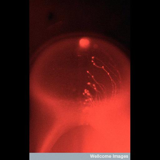
Fluorescence image of bodies and axons of the mesencephalic trigeminal nucleus in a 4 day old chick embryo. The trigeminal nucleus is made up of scattered neurons along the midline of the optic tectum. This micrograph shows the optic tectum from the back. The axons are labelled on one side only. Mesencephalic trigeminal neurons are unusual in that they are primary sensory neurons (probably derived from neural crest) that develop within the central nervous system.

Technical Details

B0001660 Cell bodies and axons. Wellcome Images available under the following creative commons usage http://creativecommons.org/licenses/by-nc-nd/2.0/uk/

Biological Sources
NCBI Organism Classification
Gallus gallus
Cell Type
sensory neuron
Cellular Component
neuron projection
neuronal cell body
Biological Context
Biological Process
mesencephalic trigeminal nucleus development
Attribution
Names
Richard Wingate
Link
Wellcome Images
Citation
Digital Object Identifier (DOI)
doi:10.7295/W9CIL38986
Archival Resource Key (ARK)
ark:/b7295/w9cil38986
Imaging
Image Type
recorded image
Image Mode
fluorescence microscopy
Parameters Imaged
fluorescence emission
Source of Contrast
distribution of a specific protein
Visualization Methods
intracellular filling
Processing History
unprocessed raw data
Sample Preparation
Relation To Intact Cell
whole mounted tissue
Dimensions
Spatial Axis Image Size Pixel Size
X 364px ——
Y 576px ——