Alternate header for print version


Licensing
Attribution Non-Commercial; No Derivatives:This image is licensed under a Creative Commons Attribution, Non-Commercial, No Derivatives License. View License Deed | View Legal Code
tweet  
*CIL – Cell Image Library accession number. Please use this to reference an image.

CIL:38994*  Cite 
Description

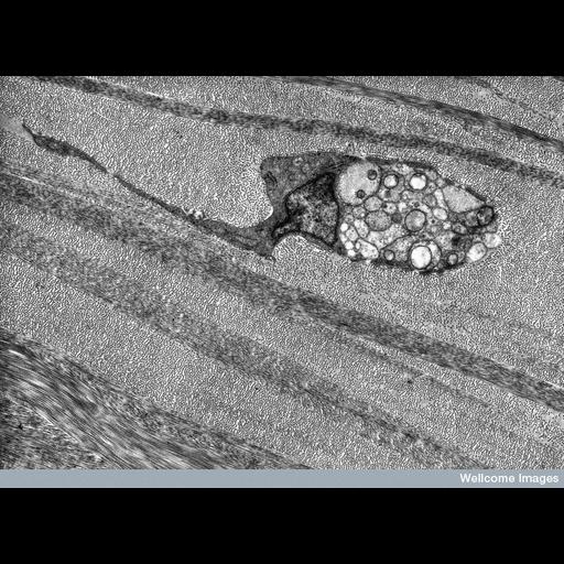
Transmission electron micrograph showing a transverse section through a nerve fiber in the corneal stroma. The cell contains a nucleus, mitochondria and some rough endoplasmic reticulum. The nerve is passing through the layers of stromal lamellae, and layers of collagen fibrils lying across each other at different angles appear as dots or lines depending on the plane of section. The field is approximately 11 microns across.

Technical Details

B0001806 Nerve fibre in corneal stroma. Wellcome Images available under the following creative commons usage http://creativecommons.org/licenses/by-nc-nd/2.0/uk/

Biological Sources
Cell Type
nerve fiber
Cellular Component
mitochondrion
rough endoplasmic reticulum
fibrillar collagen
nucleus
Biological Context
Biological Process
corneal stroma organization
Attribution
Names
Rob Young
Link
Wellcome Images
Citation
Digital Object Identifier (DOI)
doi:10.7295/W9CIL38994
Archival Resource Key (ARK)
ark:/b7295/w9cil38994
Imaging
Image Type
recorded image
Image Mode
transmission electron microscopy (TEM)
Parameters Imaged
electron density
Source of Contrast
differences in adsorption or binding of stain
Processing History
unprocessed raw data
Sample Preparation
Methods
chemically fixed tissue
Relation To Intact Cell
sectioned tissue
Dimensions
Spatial Axis Image Size Pixel Size
X 787px ——
Y 576px ——