Alternate header for print version


Licensing
Attribution Non-Commercial; No Derivatives:This image is licensed under a Creative Commons Attribution, Non-Commercial, No Derivatives License. View License Deed | View Legal Code
tweet  
*CIL – Cell Image Library accession number. Please use this to reference an image.

CIL:39026*  Cite 
Description

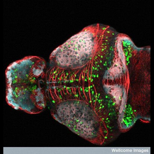
Confocal micrograph of the anterior region of the developing zebrafish brain (consisting of the telencephalon, part of diencephalon and the optic tectum). Some of the neurons (shown in green) express the green fluorescent protein (GFP) under the control of specific gene expression. Axons, tracts and neuropils have been labeled using antibodies that mark tubulin (in red) and synaptic vesicles (in blue). Tubulin is important for microtubule formation and cell structure, whereas synaptic vesicles are important for neuronal signaling. CIL 39025 is the dorsal view.

Technical Details

B0007771 Dorsal view of a zebrafish brain. Wellcome Images available under the following creative commons usage http://creativecommons.org/licenses/by-nc-nd/2.0/uk/

Biological Sources
NCBI Organism Classification
Danio rerio
Cell Type
neuron
Cellular Component
microtubule
synaptic vesicle
axon
neuropil
Biological Context
Biological Process
neuron projection development
Zebrafish Anatomy
brain
telencephalon
diencephalon
optic tectum
Attribution
Names
Monica Folgueira
Steve Wilson
Link
Wellcome Images
Citation
Digital Object Identifier (DOI)
doi:10.7295/W9CIL39026
Archival Resource Key (ARK)
ark:/b7295/w9cil39026
Grouping This image is part of a group.
Sample Preparation
Relation To Intact Cell
whole mounted tissue
Dimensions
Spatial Axis Image Size Pixel Size
X 550px ——
Y 576px ——