Alternate header for print version


Licensing
Attribution Non-Commercial; No Derivatives:This image is licensed under a Creative Commons Attribution, Non-Commercial, No Derivatives License. View License Deed | View Legal Code
tweet  
*CIL – Cell Image Library accession number. Please use this to reference an image.

CIL:39027*  Cite 
Description

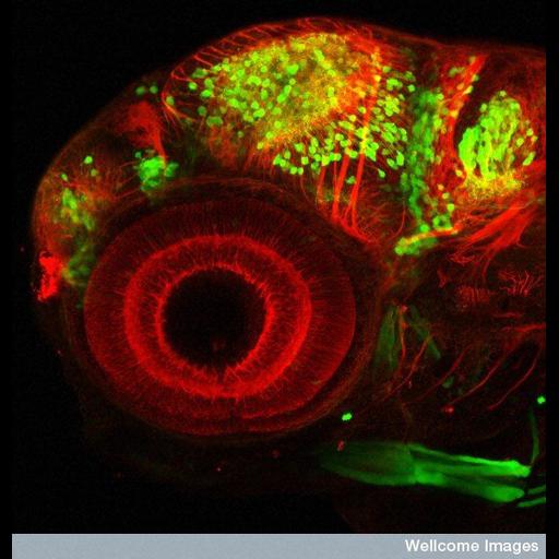
Confocal micrograph of the head region of a transgenic zebrafish embryo. The large circular structure is the eye, showing the structure of the retina. Some neurons in the brain are highlighted in green by GFP expressed under the control of a specific promoter Dlx4/6. The DLX proteins are postulated to play a role in forebrain and facial development. Therefore, the GFP is only 'switched on' in developing neurons that require this gene. The microtubules in neuronal processes have been labelled using an antibody for tubulin (red).

Technical Details

Wellcome Images available under the following creative commons usage http://creativecommons.org/licenses/by-nc-nd/2.0/uk/

Biological Sources
NCBI Organism Classification
Danio rerio
Cell Type
neuron
Cellular Component
neuron projection
neuronal cell body
microtubule
Biological Context
Biological Process
Dlx4/6 promotor specification
retina layer formation
Zebrafish Anatomy
eye
brain
Attribution
Names
Monica Folgueira
Steve Wilson
Link
Wellcome Images
Citation
Digital Object Identifier (DOI)
doi:10.7295/W9CIL39027
Archival Resource Key (ARK)
ark:/b7295/w9cil39027
Sample Preparation
Methods
chemically fixed tissue
Relation To Intact Cell
whole mounted tissue
Dimensions
Spatial Axis Image Size Pixel Size
X 550px ——
Y 576px ——