Alternate header for print version


Licensing
Attribution Non-Commercial; No Derivatives:This image is licensed under a Creative Commons Attribution, Non-Commercial, No Derivatives License. View License Deed | View Legal Code
tweet  
*CIL – Cell Image Library accession number. Please use this to reference an image.

CIL:39056*  Cite 
Description

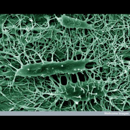
This colorized scanning electron micrograph shows the structure of osteocyte cells in the cortex of a mouse tibia bone. Osteocytes are the cells that form new bone. Imbedding the bone in resin, which was subsequently etched with perchloric acid, created this particular image by removing the entire mineral in the sample leaving a replica of the area. Therefore, what is observed is the resin that filled the spaces in the bone and the spaces inside the cells.

Technical Details

B0007595 Osteocytes. Wellcome Images available under the following creative commons usage http://creativecommons.org/licenses/by-nc-nd/2.0/uk/

Biological Sources
NCBI Organism Classification
Mus musculus
Cell Type
osteocyte
Cellular Component
cell surface
Biological Context
Biological Process
bone morphogenesis
Attribution
Names
Kevin MacKenzie
Link
wellcome images
OTHER
University of Aberdeen
Citation
Digital Object Identifier (DOI)
doi:10.7295/W9CIL39056
Archival Resource Key (ARK)
ark:/b7295/w9cil39056
Imaging
Image Type
recorded image
Image Mode
scanning electron microscopy (SEM)
Parameters Imaged
elastic scattering of electrons
Source of Contrast
differences in deposition of metal shadow
Visualization Methods
visualization of contiguous regions
Processing History
colorized
Sample Preparation
Methods
resin embedment
Relation To Intact Cell
etch
Dimensions
Spatial Axis Image Size Pixel Size
X 734px ——
Y 576px ——