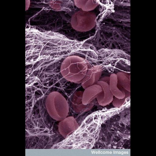
Colorized scanning electron micrograph of the components of a blood clot. Clotting of the blood is an important process that helps the body repair after injury. The blood is composed of red blood cells, which carry oxygen (shown in red), white blood cells that help fight infection (not present in this image) and platelets that are crucial in the clotting process. Platelets travel to the injured area to form a plug. Once there, they are involved in activating clotting factors, the main one being fibrin. Fibrin is a protein that crosslinks with itself to form a mesh that makes up the final blood clot (shown in this image as a string-like mesh). A blood clot is also known as a thrombus and if a thrombus occurs when it is not required it can have serious medical consequences, such as a stroke or heart attack.
B0007588 Blood clot. Wellcome Images available under the following creative commons usage http://creativecommons.org/licenses/by-nc-nd/2.0/uk/
| Spatial Axis | Image Size | Pixel Size |
|---|---|---|
| X | 389px | —— |
| Y | 576px | —— |