Alternate header for print version


Licensing
Attribution Non-Commercial; No Derivatives:This image is licensed under a Creative Commons Attribution, Non-Commercial, No Derivatives License. View License Deed | View Legal Code
tweet  
*CIL – Cell Image Library accession number. Please use this to reference an image.

CIL:39063*  Cite 
Description

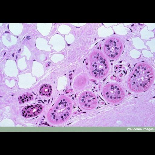
Light micrograph of a 'semithin' section (1.5 µm thick) of the dermis layer of skin from a monkey. The upper left side shows an area of (empty) fat cells surrounded by collagen fibers. The lower right side contains six sections of a coiled sweat gland and three excretory ducts. The columnar cells forming the wall of the sweat gland secrete the solutes, these are interdispersed with myosecretory cells (triangular red shapes particularly visible in gland at far right) that can contract to squeeze the secretion out of the lumen of the gland into the excretory duct. The section was stained with haematoxylin and eosin (H+E), which is a common histological stain added to tissue sections to distinguish cells.

Technical Details

B0007566 Primate sweat glands. Wellcome Images available under the following creative commons usage http://creativecommons.org/licenses/by-nc-nd/2.0/uk/

Biological Sources
Cell Type
fat cell
myosecretory cells
Cellular Component
fibrillar collagen
Biological Context
Biological Process
sweat gland organization
Human Development Anatomy
sweat gland
excretory component
skin
Attribution
Names
Spike Walker
Link
Wellcome Images
Citation
Digital Object Identifier (DOI)
doi:10.7295/W9CIL39063
Archival Resource Key (ARK)
ark:/b7295/w9cil39063
Imaging
Image Type
recorded image
Image Mode
bright-field microscopy
Parameters Imaged
absorption of illumination
Source of Contrast
differences in adsorption or binding of stain
Visualization Methods
hematoxylin
eosin
Processing History
unprocessed raw data
Sample Preparation
Methods
chemically fixed tissue
Relation To Intact Cell
sectioned tissue
Dimensions
Spatial Axis Image Size Pixel Size
X 800px ——
Y 561px ——