Alternate header for print version


Licensing
Attribution Non-Commercial; No Derivatives:This image is licensed under a Creative Commons Attribution, Non-Commercial, No Derivatives License. View License Deed | View Legal Code
tweet  
*CIL – Cell Image Library accession number. Please use this to reference an image.

CIL:39064*  Cite 
Description

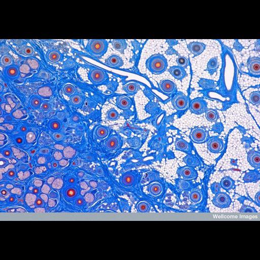
Light micrograph of a section cut through the dermis of the scalp. The dermis is the lower (or innermost) layer of the that makes up the skin; the other layer is the epidermis. The dermis contains hair follicles, sweat glands, sebaceous glands, blood vessels and lymph vessels. The section is cut tangentially in a plane +/- parallel to the skin surface. Hair follicles (blue and bright red circles) are seen in transverse sections on the left hand side, sebaceous glands are shown as pink masses ,and groups of sweat ducts are seen as patches of small dark blue circles, all of which are embedded in a matrix of blue collagen fibers. The right hand side is dominated by adipose tissue (fat), as white spaces among which are scattered hair follicles and groups of sweat glands.

Technical Details

B0007564 Scalp. Wellcome Images available under the following creative commons usage http://creativecommons.org/licenses/by-nc-nd/2.0/uk/

Biological Sources
Cell Type
hair follicle cell
sweat secreting cell
Cellular Component
fibrillar collagen
Biological Context
Biological Process
scalp dermis organization
Human Development Anatomy
dermis
sebaceus glands
adipose tissue
Attribution
Names
Spike Walker
Link
Wellcome Images
Citation
Digital Object Identifier (DOI)
doi:10.7295/W9CIL39064
Archival Resource Key (ARK)
ark:/b7295/w9cil39064
Imaging
Image Type
recorded image
Image Mode
modes of light microscopy
Parameters Imaged
absorption of illumination
Source of Contrast
differences in adsorption or binding of stain
Processing History
unprocessed raw data
Sample Preparation
Relation To Intact Cell
sectioned tissue
Dimensions
Spatial Axis Image Size Pixel Size
X 800px ——
Y 560px ——