Alternate header for print version


Licensing
Attribution Non-Commercial; No Derivatives:This image is licensed under a Creative Commons Attribution, Non-Commercial, No Derivatives License. View License Deed | View Legal Code
tweet  
*CIL – Cell Image Library accession number. Please use this to reference an image.

CIL:39110*  Cite 
Description

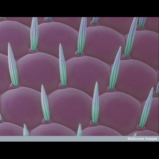
Scanning electron micrograph of the Drosophila compound eye. Close-up shows the multi-faceted cornea (pink) and the thin, hair-like structures called setae (green) which are believed to reduce glare.

Technical Details

B0007196 Drosophila eye. Wellcome Images available under the following creative commons usage http://creativecommons.org/licenses/by-nc-nd/2.0/uk/

Biological Sources
NCBI Organism Classification
Drosophila
Cell Type
cornea cell
setae
Biological Context
Biological Process
compound eye development
Attribution
Names
Anne Weston
Link
Wellcome Images
OTHER
LRI, CRUK
Citation
Digital Object Identifier (DOI)
doi:10.7295/W9CIL39110
Archival Resource Key (ARK)
ark:/b7295/w9cil39110
Imaging
Image Type
recorded image
Image Mode
scanning electron microscopy (SEM)
Parameters Imaged
elastic scattering of electrons
Source of Contrast
differences in deposition of metal shadow
Processing History
colorized
Sample Preparation
Methods
chemically fixed tissue
Relation To Intact Cell
whole mounted tissue
Dimensions
Spatial Axis Image Size Pixel Size
X 734px ——
Y 576px ——