Alternate header for print version


Licensing
Public Domain: This image is in the public domain and thus free of any copyright restrictions. However, as is the norm in scientific publishing and as a matter of courtesy, any user should credit the content provider for any public or private use of this image whenever possible. Learn more
tweet  
*CIL – Cell Image Library accession number. Please use this to reference an image.

CIL:39463*  Cite 
Description

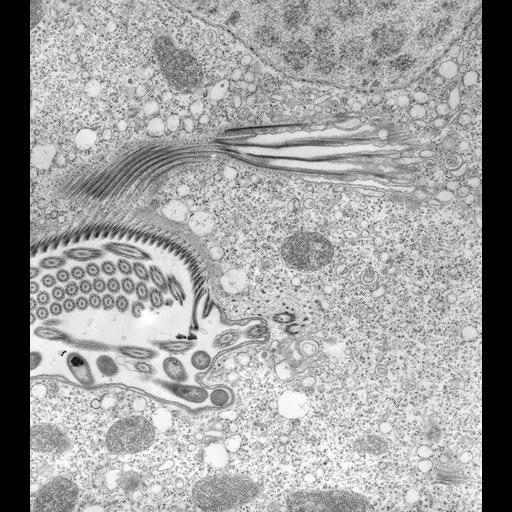
Food vacuoles arise from the single membrane that lines the cytopharynx. Vesicles, similar to discoidal vesicles, attach to the cytopharyngeal microtubules (lamellar clumps) that arise from the dorsal ribbed wall of the peristome and that pass beyond the cytosome. The cytopharynx swells and a food vacuole balloons out through the ribs that are produced by the folded cytopharyngeal membrane and the attached lamellae. How, why and when a food vacuole pinches off needs further study. A myoneme equivalent to the cytostomal cord in Paramecium lies along the origins of the lamellae (called cytopharyngeal ribbons in Paramecium). TEM taken on 3/30/71 by R. Allen with Hitachi HU11A operating at 75kV. Mag. 9,250X. The raw negative was scanned with an Epson Perfection V750 Pro and this high resolution image is best used for quantitative analysis. Additional information available at (http://www5.pbrc.hawaii.edu/allen/).

Technical Details

Standard glutaraldehyde fixation followed by osmium tetroxide, dehydrated in alcohol and embedded in an epoxy resin. Microtome sections prepared at approximately 75nm thickness. Additional information available at (http://www5.pbrc.hawaii.edu/allen/).

Biological Sources
NCBI Organism Classification
Vorticella convallaria
Cell Type
cell by organism
eukaryotic cell
Eukaryotic Protist
Ciliated Protist
Cell Line
Cambridge Culture Collection, Cambridge, England, # LB1690/1
Cellular Component
oral apparatus
food vacuole
cortical microtubule cytoskeleton
Biological Context
Biological Process
response to food
digestive system process
Attribution
Names
Richard Allen (University of Hawaii)
Citation
Digital Object Identifier (DOI)
doi:10.7295/W9CIL39463
Archival Resource Key (ARK)
ark:/b7295/w9cil39463
Grouping This image is part of a group.
Imaging
Image Type
transmission electron microscopy (TEM)
illumination by electrons
Image Mode
detection of electrons
film
Parameters Imaged
electron density
Source of Contrast
stain with broad specificity
Visualization Methods
stain with broad specificity
osmium tetroxide
uranyl salt
lead salt
Processing History
recorded image
film
Data Qualifiers
raw, unprocessed data
suitable for spatial measurements
Dimensions
Spatial Axis Image Size Pixel Size
X 3537px 1.1nm
Y 4000px 1.1nm