Alternate header for print version


Licensing
Attribution Non-Commercial; No Derivatives:This image is licensed under a Creative Commons Attribution, Non-Commercial, No Derivatives License. View License Deed | View Legal Code
tweet  
*CIL – Cell Image Library accession number. Please use this to reference an image.

CIL:39465*  Cite 
Description

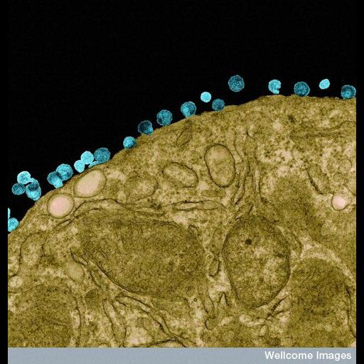
Colorized transmission electron micrograph of human immunodeficiency virus (HIV) particles (blue) budding from the surface of a T cell (a type of lymphocyte).The viruses replicate inside the cell with the different components gathering at the cell membrane to be assembled into new virus particles.

Technical Details

B0005750 HIV particles budding from a lymphocyte . Wellcome Images available under the following creative commons usage http://creativecommons.org/licenses/by-nc-nd/2.0/uk/

Biological Sources
Cell Type
lymphocyte
human immunodeficiency virus
Biological Context
Biological Process
HIV budding from lymphocyte
Attribution
Names
R. Dourmashkin
Link
Wellcome Images
Citation
Digital Object Identifier (DOI)
doi:10.7295/W9CIL39465
Archival Resource Key (ARK)
ark:/b7295/w9cil39465
Imaging
Image Type
recorded image
Image Mode
transmission electron microscopy (TEM)
Parameters Imaged
detection of electrons
Source of Contrast
differences in adsorption or binding of stain
Processing History
colorized
Sample Preparation
Relation To Intact Cell
sectioned tissue
Dimensions
Spatial Axis Image Size Pixel Size
X 522px ——
Y 576px ——