Alternate header for print version


Licensing
Attribution Non-Commercial Share Alike:This image is licensed under a Creative Commons Attribution, Non-Commercial Share Alike License. View License Deed | View Legal Code
tweet  
*CIL – Cell Image Library accession number. Please use this to reference an image.

CIL:39731*  Cite 
Description

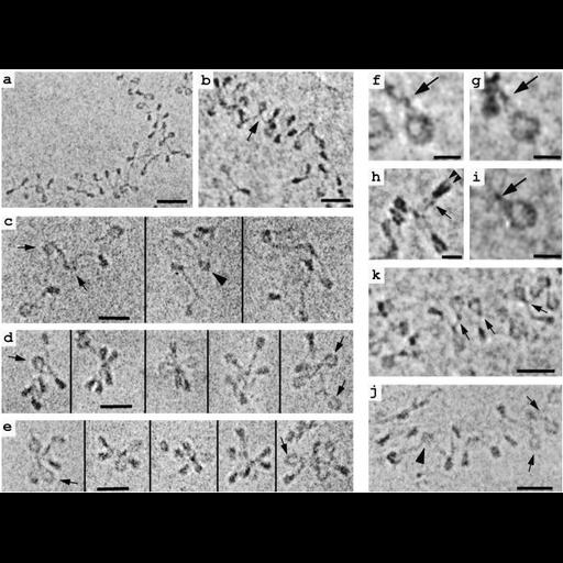
TEM image of frozen hydrated soluble chromatin isolated from chicken erythrocyte nuclei and vitrified in low salt. Polynucleosomes adopt an irregular zigzag fiber conformation (a,b,k), and show a 'stem' motif. Similar features are seen in reconstituted polynucleosomes (c,d,f-i). However, if reconstitutes lack linker histone H5, the stem conformation is absent (c). See Fig 1 in J Bednar et al. 1998 Nucleosomes, linker DNA and linker histone form a unique structural motif that directs the higher-order folding and compaction of chromatin. Proc Natl Acad Sci USA 95:14173-14178.

Technical Details

Isolated nuclei were prepared from chicken erythrocyte nuclei and polynucleosomes released after treatment with micrococcal nuclease. Reconstituted polynucleosomes were prepared by salt dialysis using purified DNA and histones. Samples were fixed with 0.1% glutaraldehyde for 4 hr at 4C, placed on holey carbon films, vitrified by plunge freezing, and observed in the frozen hydrated state using a Philips CM10 TEM operated at 60KV. Low dose images were recorded on Kodak SO163 film. See also: J Bednar et al. 1998 Nucleosomes, linker DNA and linker histone form a un ique structural motif that directs the higher-order folding and compaction of chromatin. Proc Natl Acad Sci USA 95:14173-14178.

Biological Sources
NCBI Organism Classification
Gallus gallus gallus
Cell Type
erythrocyte
Cellular Component
chromatin
nucleosome
heterochromatin
Attribution
Names
Jan Bednar
Rachel Horowitz
Sergei Grigoryev
Lenny Carruthers
Jeffrey Hansen
Abraham Koster
Christopher Woodcock
Published
J Bednar et al. 1998 Nucleosomes, linker DNA and linker histone form a unique structural motif that directs the higher-order folding and compaction of chromatin. Proc Natl Acad Sci USA 95:14173-14178.
Pubmed
9826673
Link
Proceedings National Academy of Sciences USA
Citation
Digital Object Identifier (DOI)
doi:10.7295/W9CIL39731
Archival Resource Key (ARK)
ark:/b7295/w9cil39731
Grouping This image is part of a group.
Imaging
Image Type
film
Image Mode
transmission electron microscopy (TEM)
Parameters Imaged
electron density
Source of Contrast
intrinsic mass distribution
Visualization Methods
visualization of contiguous regions
Data Qualifiers
processed data
Sample Preparation
Methods
glutaraldehyde fixed tissue
cryofixed tissue
Relation To Intact Cell
isolated subcellular component
Dimensions
Spatial Axis Image Size Pixel Size
X 675px ——
Y 508px ——