Alternate header for print version


Licensing
Attribution Only: This image is licensed under a Creative Commons Attribution License. View License Deed | View Legal Code
tweet  
*CIL – Cell Image Library accession number. Please use this to reference an image.

CIL:39949*  Cite 
Description

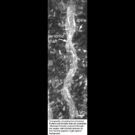
Tomographic reconstruction of a dendrite and dendritic spines from a Purkinje neuron in the rat cerebellum. This image is a maximum intensity projection through the volume of a segment of dendrite in a 2 um section, with contrast reversed so that dendrite appears bright against background. This reconstructed image has been downsampled from the raw data image, which can be accessed using the link provided to the Cell Centered Database.

Technical Details

The tomogram was generated using an JEOL4000 IVEM. Single tilt images spanned -60 to 60° in 2° increments. Magnification, 3000X; accelerating voltage, 400.0 KeV.

Biological Sources
NCBI Organism Classification
Rattus norvegicus
Cell Type
CNS neuron (sensu Vertebrata)
Purkinje cell
Cellular Component
dendritic spine
dendrite
Attribution
Names
Maryann Martone
Naoko Yamada
Gordun Arbuthnott
Cali Ingham
Stephen Young
Link
CCDB 13
Citation
Digital Object Identifier (DOI)
doi:10.7295/W9CIL39949
Archival Resource Key (ARK)
ark:/b7295/w9cil39949
Imaging
Image Type
recorded image
Image Mode
transmission electron microscopy (TEM)
detection of electrons
illumination by electrons
Parameters Imaged
electron density
Data Qualifiers
processed data
Sample Preparation
Relation To Intact Cell
microtome-sectioned tissue
Dimensions
Spatial Axis Image Size Pixel Size
X 171px ——
Y 583px ——