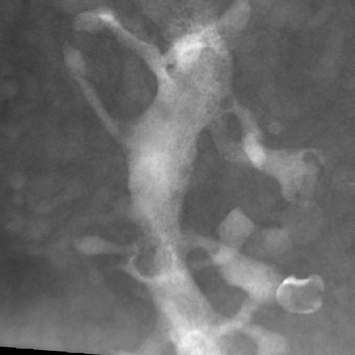
Tomographic reconstruction of dendritic spines on a dendrite from a medium spiny neuron from the rat neostriatum. This image is a maximum intensity projection of a segment of dendrite in a 2 um section. This reconstructed image has been downsampled from the raw data image, which can be accessed using the link provided to the Cell Centered Database.
The tomogram was generated using an Hitachi 1MeV HVEM. Single tilt images spanned -60 to 60° in 2° increments. Magnification, 10000.0 X; accelerating voltage, 1.0 MeV.
| Spatial Axis | Image Size | Pixel Size |
|---|---|---|
| X | 512px | —— |
| Y | 512px | —— |