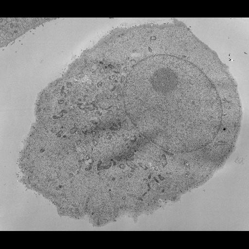
Mosaic electron micrograph covering the entire cross section of a VeroE6 cell infected with SARS-CoV. The image represents a 100nm thick plane through the center of the infected cell. This image has been downsampled from the raw data image which can be accessed using the link provided to the Cell Centered Database. For more information, see: Knoops et al. SARS-coronavirus replication is supported by a reticulovesicular network of modified endoplasmic reticulum. PLoS Biol. 2008 6(9):e226. PMID: 18798692
For ultrastructural morphological investigations, SARS-CoV-infected Vero E6 cells were pre-fixed (for biosafety reasons) overnight with 3% paraformaldehyde in 0.1M PHEM buffer (60 mM piperazide-1,4-bis[2-ethanesulfonic acid], 25 mM HEPES, 2 mM MgCl2, 10 mM EGTA) at various time points after infection. For cryo-fixation, cell monolayers adhered to Thermanox coverslips (Nunc, Denmark) were plunged into liquid ethane. Freeze substitution was performed at -90°C in an automated freeze-substitution system (Leica, Austria) using an FS medium consisting of 90% acetone and 10% water, containing 1% osmium tetroxide and 0.5 % uranyl acetate. After washing with pure acetone at RT, the samples were embedded in epoxy LX-12 resin. Thin sections were collected on 100 Mesh grids, 1.25% pioloform support film contrasted with uranyl acetate and lead hydroxide and subsequently viewed at 80 kV with a Philips CM-10 transmission electron microscope (Philips, The Netherlands).
| Spatial Axis | Image Size | Pixel Size |
|---|---|---|
| X | 512px | —— |
| X | 512px | —— |