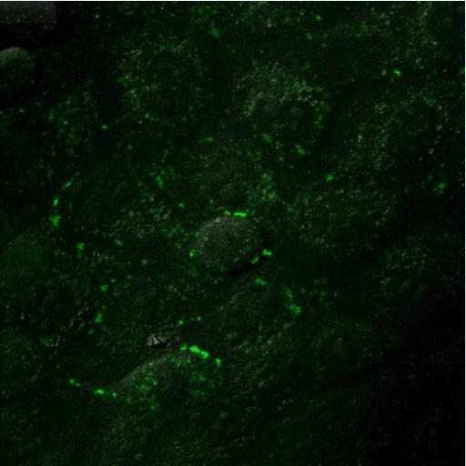
A single frame from a time-lapse animation of FlAsH-labeled MDCK cells expressing Cx43-4C309/337 (green) showing their rearrangement and fate during and after mitosis. In the time-lapse series, which can be accessed using the link provided to the Cell Centered Database, FlAsH fluorescence excitation z series along with a transmitted DIC image were recorded every 6 min for a total duration of about 5 h. 3D volume reconstructions from confocal image stacks over time are displayed at a rate of two frames per minute. From Supplemental movie 2 from Boassa et al. (2010) Traffic. PMID 20716111.
MDCK cells stably expressing two internal 4C domains in the C-terminus (FLNCCPGCCME)-tagged Cx43 (Cx43-4C309/337) were labeled for 1 h at 37 degrees C with 180 nM FlAsH-EDT2/12.5 uM EDT in Hanks balanced salt saline (HBSS). Free and non-specifically bound FlAsH was removed by washing with 2,3-dimercapto-1-propanol (BAL, 500 uM, 20min at 37 degrees C in HBSS). Time-lapse imaging was conducted using an Olympus FluoView1000 confocal microscope equipped with a temperature controlled chamber (at 37 degrees C) and a 60X 1.42 NA objective. Medium: Opti-MEM supplemented with 5% FBS covered with a glass coverslip.
| Spatial Axis | Image Size | Pixel Size |
|---|---|---|
| X | 512px | —— |
| X | 512px | —— |