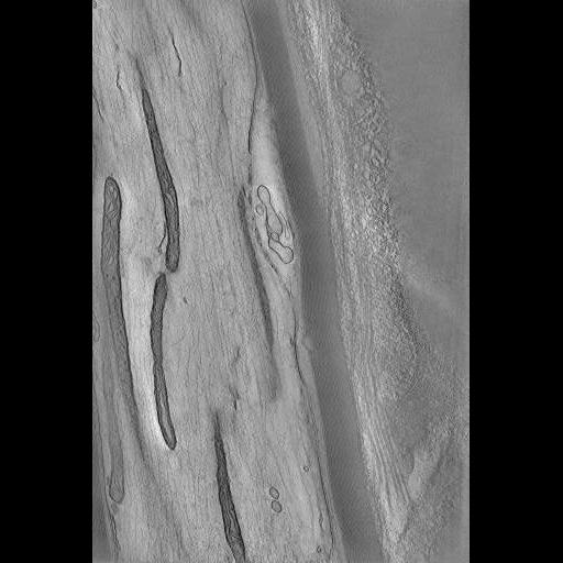
A single image from animation of the computed slices through a tomographic volume of mitochondria at the Node of Ranvier in rat peripheral nerve root. This image has been downsampled from the raw data image which can be accessed using the link provided to the Cell Centered Database. For more information, see: Perkins et al. Mitochondrial configurations in peripheral nerve suggest differential ATP production. J. Struct. Biol. (2010), doi:10.1016/j.jsb.2010.06.017 http://www.ncbi.nlm.nih.gov/pubmed/20600951
Chemical fixation with 2% paraformaldehyde and 2.5% glutaraldehyde via vascular perfusion in 0.15M cacodylate buffer (pH 7.4) at 37 degrees C followed by high pressure freezing and freeze substitution using a Bal Tec HPM010 high pressure freezer (from Perkins et al., J. Structural Biology 161, 469, 2008).