Alternate header for print version


Licensing
Public Domain: This image is in the public domain and thus free of any copyright restrictions. However, as is the norm in scientific publishing and as a matter of courtesy, any user should credit the content provider for any public or private use of this image whenever possible. Learn more
tweet  
*CIL – Cell Image Library accession number. Please use this to reference an image.

CIL:40379*  Cite 
Description

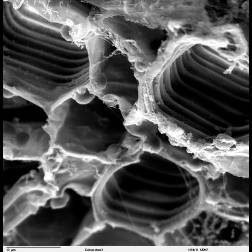
Scanning electron microscope image of cross-section through a Solenostemon scutellarioidesi (coleus) stem. This image is a high magnification view of CIL 40378 showing x-section through the vascular bundles. This image is also part of a group on botanical stems (CIL:40378-40395).

Technical Details

Image collected on a Zeiss DSM 962 SEM. Complete specimen preparation protocol available at: http://remf.dartmouth.edu:8080/EM-Wiki/36

Biological Sources
NCBI Organism Classification
Solenostemon scutellarioides
Biological Context
Biological Process
vascular bundle organization
Attribution
Names
Louisa Howard
Link
Dartmouth College EM Facility
Citation
Digital Object Identifier (DOI)
doi:10.7295/W9CIL40379
Archival Resource Key (ARK)
ark:/b7295/w9cil40379
Grouping This image is part of a group.
Sample Preparation
Methods
glutaraldehyde fixed tissue
ethanol dehydration
critical_point dried specimen
Relation To Intact Cell
sectioned tissue
Dimensions
Spatial Axis Image Size Pixel Size
X 1024px ——
Y 1049px ——