Alternate header for print version


Licensing
Attribution Non-Commercial Share Alike:This image is licensed under a Creative Commons Attribution, Non-Commercial Share Alike License. View License Deed | View Legal Code
tweet  
*CIL – Cell Image Library accession number. Please use this to reference an image.

CIL:40510*  Cite 
Description

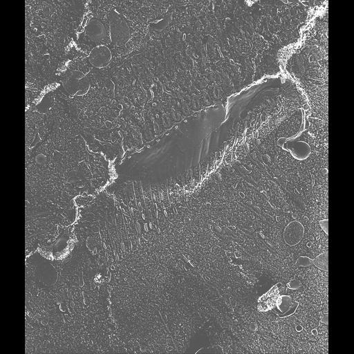
Quick-freeze deep-etch rotary-shadowed replica of a portion of the chemically-unfixed cytopharynx. The cytopharynx is continuous with the plasma membrane and is covered by only one membrane which is pleated over the ends of the cytopharyngeal microtubular ribbons. Numerous discoidal vesicles are bound single file along the anterior side only of each ribbon. This is the site where discoidal vesicles fuse with the cytopharyngeal membrane to form the digestive vacuole. TEM taken on 5/18/88 by C. Schroeder with Zeiss 10A operating at 80kV. Neg. 6,300X. The raw negative was scanned with an Epson Perfection V750 Pro and this high resolution image is best used for quantitative analysis. Additional information available at (http://www5.pbrc.hawaii.edu/allen/). Part published in J. Cell Biol. 111:2553-2562, 1990. Adapted with permission.

Biological Sources
NCBI Organism Classification
Paramecium multimicronucleatum
Cell Type
cell by organism
eukaryotic cell
Eukaryotic Protist
Ciliated Protist
Cellular Component
oral apparatus
microtubule associated complex
Biological Context
Biological Process
oral apparatus organization
Attribution
Names
Richard Allen (University of Hawaii)
C. Schroeder
Published
J Cell Biol. 1990 Dec;111(6 Pt 1):2553-62.
Pubmed
2148940
Citation
Digital Object Identifier (DOI)
doi:10.7295/W9CIL40510
Archival Resource Key (ARK)
ark:/b7295/w9cil40510
Grouping This image is part of a group.
Imaging
Image Type
transmission electron microscopy (TEM)
illumination by electrons
Image Mode
electron density
Parameters Imaged
elastic scattering of electrons
electron density
elevation
Source of Contrast
differences in deposition of metal shadow
Visualization Methods
transmission electron microscopy (TEM)
Processing History
freeze_fracture/freeze_etch
shadowing and plating
recorded image
film
Data Qualifiers
raw, unprocessed data
suitable for spatial measurements
Dimensions
Spatial Axis Image Size Pixel Size
X 3432px 1.6nm
Y 4000px 1.6nm