Alternate header for print version


Licensing
This image is copyright protected. Any public or private use of this image is subject to prevailing copyright laws. Please contact the content provider of this image for permission requests.
tweet  
*CIL – Cell Image Library accession number. Please use this to reference an image.

CIL:40593*  Cite 
Description

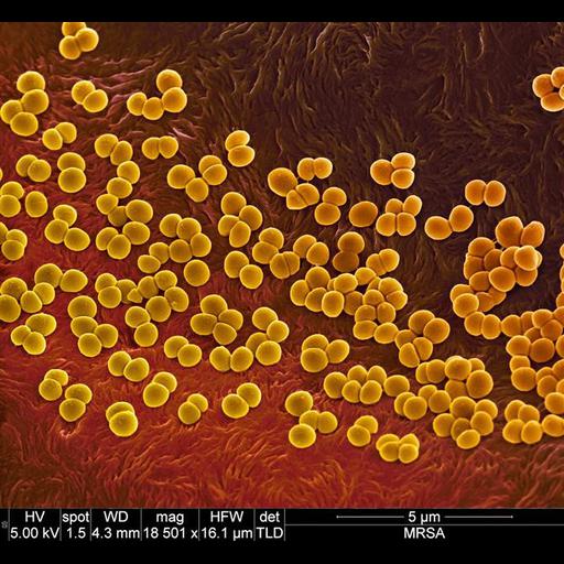
Colorized scanning electron micrograph of methicillin resistant staphylococcus aureus (MRSA) bacteria on the surface of a wound dressing.

Technical Details

Specimen was fixed with glutaraldehyde and dehydrated through an ethanol series. It was coated with 5nm gold-palladium and imaged at ultra high resolution (semi-immersion optics). The image was collected on a FEI Nova NanoSEM Family with the following imaging parameters: Magnification: 18,501 x, Horizontal Field Width: 16.1 microns, Vacuum: High vacuum (approx 10 e-06 Torr), Voltage: 5kV, Detector: TLD, Spot: 1.5 nA, and Working Distance: 4.3 mm

Biological Sources
NCBI Organism Classification
Staphylococcus aureus
Cellular Component
cell surface
Biological Context
Biological Process
antibiotic resistance
Attribution
Names
Paul Gunning
Link
FEI Life Sciences Images
Citation
Digital Object Identifier (DOI)
doi:10.7295/W9CIL40593
Archival Resource Key (ARK)
ark:/b7295/w9cil40593
Imaging
Image Type
recorded image
Image Mode
scanning electron microscopy (SEM)
Parameters Imaged
elastic scattering of electrons
Source of Contrast
differences in deposition of metal shadow
Visualization Methods
visualization of contiguous regions
shadowing and plating
gold
palladium
Processing History
colorized
Sample Preparation
Methods
glutaraldehyde fixed tissue
ethanol dehydration
Dimensions
Spatial Axis Image Size Pixel Size
X 670px ——
Y 617px ——