Alternate header for print version


Licensing
This image is copyright protected. Any public or private use of this image is subject to prevailing copyright laws. Please contact the content provider of this image for permission requests.
tweet  
*CIL – Cell Image Library accession number. Please use this to reference an image.

CIL:40607*  Cite 
Description

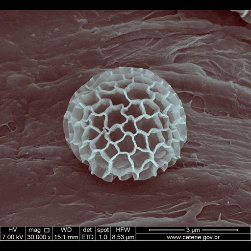
Scanning electron micrograph of a myxomycetes (a type of slime mold). This image was collected as part of a study to assess the diversity of myxomycetes in the Atlantic Forest.

Technical Details

FEI instrument: Quanta Family Magnification: 30,000x Horizontal Field Width: 8.53 μm Voltage: 7 kV Detector: ETD Spot: 1.0 nA Working Distance: 15.1 mm

Biological Sources
NCBI Organism Classification
Myxogastria
Cellular Component
cell surface
Attribution
Names
FRANCISCO RANGEL
Link
FEI Life Science Images
Citation
Digital Object Identifier (DOI)
doi:10.7295/W9CIL40607
Archival Resource Key (ARK)
ark:/b7295/w9cil40607
Imaging
Image Type
recorded image
Image Mode
scanning electron microscopy (SEM)
Parameters Imaged
elastic scattering of electrons
Source of Contrast
differences in deposition of metal shadow
Visualization Methods
visualization of contiguous regions
Processing History
colorized
Sample Preparation
Relation To Intact Cell
whole mounted tissue
Dimensions
Spatial Axis Image Size Pixel Size
X 670px ——
Y 617px ——