Alternate header for print version


Licensing
This image is copyright protected. Any public or private use of this image is subject to prevailing copyright laws. Please contact the content provider of this image for permission requests.
tweet  
*CIL – Cell Image Library accession number. Please use this to reference an image.

CIL:40608*  Cite 
Description

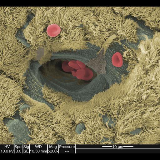
Colorized scanning electron micrograph of a mouse trachea and its red blood cells.

Technical Details

Image collected on a FEI instrument: Quanta Family using the following parameters: Magnification: 5200x, Voltage: 10.0 kV, Detector: SE, Spot: 3.0 nA, and Working Distance: 10.50 mm.

Biological Sources
NCBI Organism Classification
Mus musculus
Cell Type
erythrocyte
ciliated epithelial cell
Cellular Component
cell surface
cilium
Biological Context
Mouse Gross Anatomy
TS25,trachea
Attribution
Names
Matthew Sharp
Link
FEI Life Science Images
Citation
Digital Object Identifier (DOI)
doi:10.7295/W9CIL40608
Archival Resource Key (ARK)
ark:/b7295/w9cil40608
Imaging
Image Type
recorded image
Image Mode
scanning electron microscopy (SEM)
Parameters Imaged
elastic scattering of electrons
Visualization Methods
visualization of contiguous regions
Processing History
colorized
Sample Preparation
Methods
chemically fixed tissue
Relation To Intact Cell
sectioned tissue
Dimensions
Spatial Axis Image Size Pixel Size
X 670px ——
Y 616px ——