Alternate header for print version


Licensing
Attribution Non-Commercial Share Alike:This image is licensed under a Creative Commons Attribution, Non-Commercial Share Alike License. View License Deed | View Legal Code
tweet  
*CIL – Cell Image Library accession number. Please use this to reference an image.

CIL:40666*  Cite 
Description

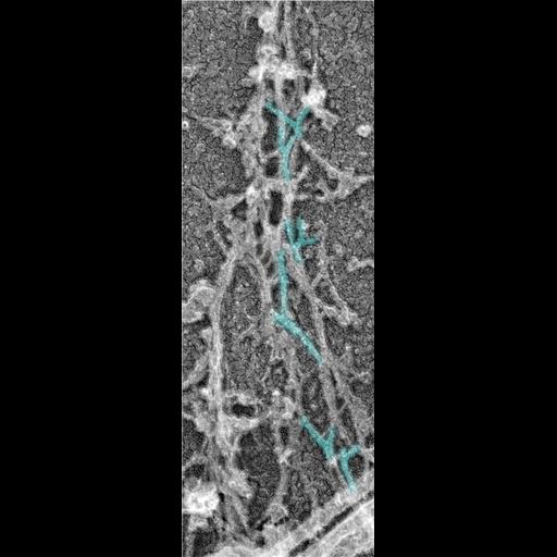
Platinum replica illustrating the cytoskeletal organization of dendritic spines from extracted 14 DIV neurons. This image shows branched actin filaments (cyan) in the neck of respective spines. This image is part of a group of images representing Figure 2B and its components from Mol Biol Cell. 2010 Jan 1;21(1):165-76. Figure 2B appears as CIL 40664. The uncolorized overview panel is CIL 40667. Box 4 appears as CIL 40665, and Box 5 (this micrograph) appears as CIL 40666.

Technical Details

Please refer to referenced article for details on cell extraction and processing for electron microscopy.

Biological Sources
NCBI Organism Classification
Rattus
Cell Type
hippocampal neuron
Cellular Component
dendritic spine
dendritic shaft
actin cytoskeleton
Biological Context
Biological Process
dendritic spine organization
dendrite morphogenesis
Attribution
Names
Farida Korobova
Tatyana Svitkina
Published
Mol Biol Cell. 2010 Jan 1;21(1):165-76. Epub 2009 Nov 4.
Pubmed
19889835
Citation
Digital Object Identifier (DOI)
doi:10.7295/W9CIL40666
Archival Resource Key (ARK)
ark:/b7295/w9cil40666
Grouping This image is part of a group.
Imaging
Image Type
recorded image
Image Mode
transmission electron microscopy (TEM)
Parameters Imaged
electron density
Source of Contrast
differences in deposition of metal shadow
Visualization Methods
shadowing and plating
Processing History
colorized
Dimensions
Spatial Axis Image Size Pixel Size
X 380px ——
Y 1096px ——