Alternate header for print version


Licensing
Attribution Non-Commercial Share Alike:This image is licensed under a Creative Commons Attribution, Non-Commercial Share Alike License. View License Deed | View Legal Code
tweet  
*CIL – Cell Image Library accession number. Please use this to reference an image.

CIL:40990*  Cite 
Description

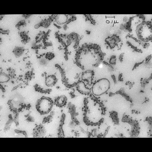
Transmission electron micrograph of a thin section of plastic-embedded material from the 'rough microsome' fraction of a preparation of rat liver. Subsequent to this early image, 'microsomes' became known as ribosomes, and the subcellular fraction consisting of membrane fragments studded with ribosomes became termed rough endoplasmic reticulum, the site of synthesis of proteins destined for export from the cell.

Technical Details

Original 3.25 in. x 4 in. lantern slides were scanned at 600dpi.

Biological Sources
NCBI Organism Classification
Rattus rattus
Cell Type
hepatocyte
Cellular Component
microsome
ribosome
rough microsome
rough endoplasmic reticulum
Biological Context
Biological Process
protein synthesis
Attribution
Names
George E. Palade
Link
George E. Palade EM Slide Collection
Citation
Digital Object Identifier (DOI)
doi:10.7295/W9CIL40990
Archival Resource Key (ARK)
ark:/b7295/w9cil40990
Grouping This image is part of a group.
Imaging
Image Type
film
Image Mode
transmission electron microscopy (TEM)
Parameters Imaged
electron density
Source of Contrast
differences in adsorption or binding of stain
Visualization Methods
uranyl salt
osmium tetroxide
Data Qualifiers
processed data
Dimensions
Spatial Axis Image Size Pixel Size
X 4156px ——
Y 3382px ——