Alternate header for print version


Licensing
This image is copyright protected. Any public or private use of this image is subject to prevailing copyright laws. Please contact the content provider of this image for permission requests.
tweet  
*CIL – Cell Image Library accession number. Please use this to reference an image.

CIL:41028*  Cite 
Description

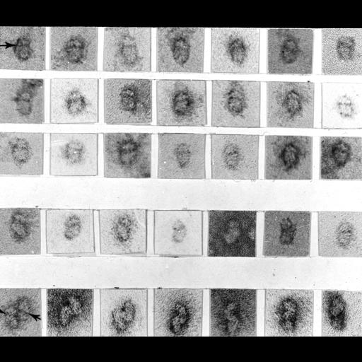
Gallery of transmission electron micrographs of ribosomes purified from a rat liver homogenate by sucrose gradient centrifugation, and negatively stained with uranyl acetate. The gallery illustrates that there is a population of particles that appears to comprise small subunits and dimers of small subunits.

Technical Details

Samples were placed on carbon coated collodion films, negatively stained with aqueous uranyl acetate, and examined with a Philips 300 TEM. See also Plate II in: N. Yoshiaki et al. 1971. Structure of liver ribosomes studied by negative staining. J Mol Biol 60:303-323

Biological Sources
NCBI Organism Classification
Rattus rattus
Cell Type
hepatocyte
Cellular Component
ribosome
Biological Context
Biological Process
protein sythesis
Attribution
Names
Yoshiaki Nonomura
Gunter Blobel
David Sabatini
George E. Palade
Published
N. Yoshiaki et al. 1971. Structure of liver ribosomes studied by negative staining. J Mol Biol 60:303-323
Pubmed
4106575
Link
George E. Palade EM Slide Collection
Citation
Digital Object Identifier (DOI)
doi:10.7295/W9CIL41028
Archival Resource Key (ARK)
ark:/b7295/w9cil41028
Grouping This image is part of a group.
Imaging
Image Type
film
Image Mode
transmission electron microscopy (TEM)
Parameters Imaged
electron density
Source of Contrast
differences in adsorption or binding of stain
Visualization Methods
uranyl salt
Data Qualifiers
processed data
Sample Preparation
Methods
unfixed tissue
Relation To Intact Cell
isolated subcellular component
Dimensions
Spatial Axis Image Size Pixel Size
X 2912px ——
Y 2503px ——