Alternate header for print version


Licensing
Attribution Non-Commercial; No Derivatives:This image is licensed under a Creative Commons Attribution, Non-Commercial, No Derivatives License. View License Deed | View Legal Code
tweet  
*CIL – Cell Image Library accession number. Please use this to reference an image.

CIL:41458*  Cite 
Description

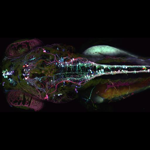
"Brainbow" zebrafish. Neurons are labeled in multiple colors with "Brainbow" (Nature, 2007) fluorescence microscopy. Three fluorescent proteins (cyan, yellow, and red) are randomly taken up by various neurons, offering a palette of dozens of colors to help scientists follow complex neural pathways. Shown here is a 5-day-old zebrafish larva viewed from the dorsal side, captured using a 20X objective. Fourth Prize, 2008 Olympus BioScapes Digital Imaging Competition®.

Technical Details

Biological Sources
NCBI Organism Classification
Danio rerio
Cell Type
neuron
Cellular Component
neuron projection
Attribution
Names
Albert Pan
2008 Olympus BioScapes Digital Imaging Competition®
Link
2008 Olympus BioScapes Digital Imaging Compe...
OTHER
Harvard University, Cambridge, MA, USA
Citation
Digital Object Identifier (DOI)
doi:10.7295/W9CIL41458
Archival Resource Key (ARK)
ark:/b7295/w9cil41458
Sample Preparation
Relation To Intact Cell
whole mounted tissue
Dimensions
Spatial Axis Image Size Pixel Size
X 1963px ——
Y 973px ——