Alternate header for print version


Licensing
Attribution Non-Commercial; No Derivatives:This image is licensed under a Creative Commons Attribution, Non-Commercial, No Derivatives License. View License Deed | View Legal Code
tweet  
*CIL – Cell Image Library accession number. Please use this to reference an image.

CIL:41624*  Cite 
Description

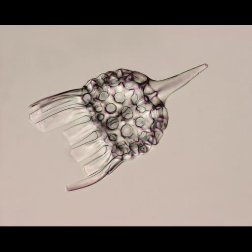
Brightfield image using an depth of fieldd showing the skeleton of a radiolarian, a single-cell protozoan with an intricate mineral skeleton. Radiolarian skeletons typically have a central capsule dividing the cell into inner and outer portions, called endoplasm and ectoplasm. Honorable Mention, 2011 Olympus BioScapes Digital Imaging Competition®.

Technical Details

Biological Sources
NCBI Organism Classification
Radiolaria
Cellular Component
skeleton
extracellular region
Biological Context
Biological Process
cytoplasmic organization
Attribution
Names
Christopher B. Jackson
2011 Olympus BioScapes Digital Imaging Competition®
Link
2011 Olympus BioScapes Digital Imaging Compet...
OTHER
Berne, Switzerland
Citation
Digital Object Identifier (DOI)
doi:10.7295/W9CIL41624
Archival Resource Key (ARK)
ark:/b7295/w9cil41624
Imaging
Image Type
recorded image
Image Mode
bright-field microscopy
extended depth of field
Parameters Imaged
absorption of illumination
Source of Contrast
differences in intrinsic optical density
Sample Preparation
Relation To Intact Cell
whole mounted tissue
Dimensions
Spatial Axis Image Size Pixel Size
X 3840px ——
Y 3072px ——